
Diagnostic Imaging: A Guide to Medical Scans
Diagnostic Imaging: A Guide to Medical Scans | The first step toward effective medical treatment always begins with an accurate diagnosis. In the medical world, the ability to “see” what is happening inside the body without invasive surgery is a technological marvel. This is what we call diagnostic imaging. Through various advanced machines and techniques, doctors can map organ structures and monitor biological activities at a cellular level to find hidden clues about a medical condition.
Understanding the Essence of Diagnostic Imaging
At its core, diagnostic imaging refers to a collection of medical procedures used to create visual representations of the interior of a body. The specific method a physician chooses typically depends on the patient’s symptoms and which part of the anatomy requires examination. This technology is not only used to detect broken bones; it is also crucial for identifying tumors, blood vessel blockages, and neurological disorders.
Each machine has unique characteristics. Some utilize sound waves, others use powerful magnetic fields, or even low-level radioactive particles. Understanding these functional differences helps patients feel more at ease when undergoing procedures at a hospital or clinic.
Common Types of Imaging Technologies
As medical specialties have evolved, imaging devices have become increasingly specialized. Here are the primary methods that serve as the pillars of modern diagnostics:
1. X-rays and CT Scans
X-rays are the oldest form of imaging but remain the most widely used, particularly for viewing bone density or detecting lung infections. A CT Scan (Computed Tomography), on the other hand, is a more sophisticated version that combines a series of X-ray images taken from different angles to create cross-sectional (3D) views of soft tissues and blood vessels.
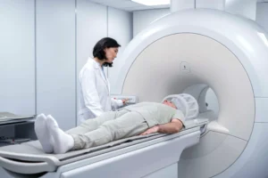
2. MRI (Magnetic Resonance Imaging)
Unlike X-rays, an MRI uses a powerful magnetic field and radio waves. This device is superior for capturing detailed images of the brain, spinal cord, and joints. Its primary advantage is that it does not expose the patient to radiation, though the procedure requires the individual to stay perfectly still for an extended period inside a tube-like machine.
3. Nuclear Medicine Scans
These procedures involve using a very small amount of a radioactive substance introduced into the body. The focus here is not just on anatomical structure, but on how the organs are actively functioning, such as in thyroid scans or the early detection of cancer spread.
4. Ultrasound
Utilizing high-frequency sound waves, ultrasound is a top choice because it is non-invasive and radiation-free. Beyond monitoring fetal development during pregnancy, ultrasound is now heavily relied upon to examine the abdomen, heart, and glands without harmful side effects.
The Patient Experience: Comfort and Specialized Procedures
Undergoing an imaging test can often trigger anxiety. In reality, the majority of exams, such as standard X-rays or ultrasounds, are quick and painless. However, some tests require extra patience. For instance, during an MRI, patients must remain calm and motionless to prevent the images from blurring. For some, staying in a narrow machine for a long time can be uncomfortable.
Additionally, there are more specific methods involving a scope. This is a tiny camera attached to a long, thin tube. A doctor moves it through natural body passageways to directly view the inside of an organ, such as the colon (colonoscopy) or lungs. Since these procedures are semi-invasive, they often require anesthesia to ensure the patient feels no pain.
Radiation and Safety Considerations
A common concern regarding imaging is radiation exposure. It is true that methods like X-rays and CT scans involve ionizing radiation in small doses. However, medical professionals always balance the diagnostic benefits against the minimal risks. Modern safety standards ensure that the amount of radiation received is kept within safe limits and is only administered when essential for treatment.
Closing the Gap of Medical Uncertainty
Ultimately, the presence of various imaging technologies aims to eliminate guesswork in medical intervention. Without clear visuals, treatment would be based on assumptions. With precise scan results, doctors can develop more targeted care plans, ranging from accurate medication dosages to better-prepared surgical procedures.
Diagnostic imaging is about more than just machines and cables; it is about providing clarity to patients regarding their health. Understanding how these tools work helps us appreciate how far science has come in preserving human quality of life.
Portable Ultrasound: Fast & Precise Diagnostics
Portable Ultrasound: Fast & Precise Diagnostics | Over the last few decades, the face of healthcare has undergone a significant shift. One of the most impactful technological leaps is the evolution of medical imaging devices. While ultrasound (USG) examinations used to require patients to visit specialized radiology rooms equipped with massive machines, the scenario has now changed. The emergence of portable ultrasound machines has broken down the hospital walls, bringing diagnostic capabilities directly to the patient’s bedside.
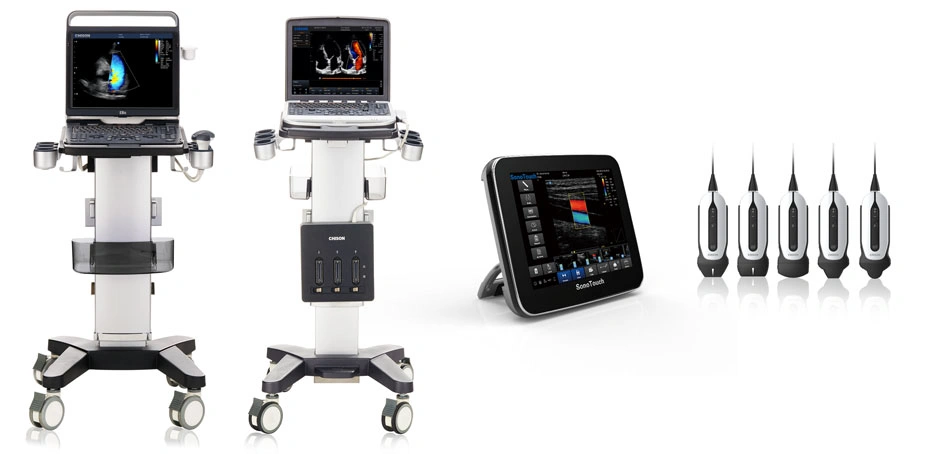
What is a Portable Ultrasound Machine?
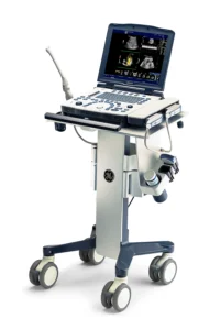
Put simply, a portable ultrasound is an imaging system designed with mobility in mind without compromising image quality. These devices no longer rely on complex wiring or heavy stationary units. In the field, they come in various forms, ranging from compact trolley models that are easy to move between rooms, to sleek tablets, and even handheld devices that fit inside a doctor’s lab coat pocket.
This technology operates on the same principles as conventional ultrasound—using high-frequency sound waves to produce real-time images of internal organs. The difference lies in its flexibility; these tools are ready for use in cramped spaces, ambulances, or even emergency tents at disaster sites.
A Catalyst for Change on the Frontlines
The rising popularity of these mobile devices did not happen by chance. The COVID-19 pandemic served as a crucial turning point that accelerated the adoption of this technology. At that time, the need for diagnostic tools that were fast, accurate, and easy to sterilize became a top priority. Portable ultrasound allowed doctors to check the lung conditions of critical patients without having to move them to a radiology suite, which simultaneously minimized the risk of virus transmission.
Over time, these benefits expanded across various departments. Emergency Departments (ED) and Intensive Care Units (ICU) were at the forefront of experiencing the efficiency of these tools. In critical situations where every second counts, clinicians can immediately perform triage and determine medical actions within minutes of a patient’s arrival.
Breaking Through Medical Specialties
The ability to move between departments has made portable ultrasound a “modern stethoscope” for many specialists. Its use is no longer limited to monitoring pregnancies in OB/GYN clinics; it has branched out into various other fields:
-
Cardiology & Vascular: Instantly monitoring blood flow and heart conditions.
-
Endocrinology: Facilitating precise ultrasound-guided thyroid biopsies.
-
Pediatrics: Providing a less intimidating examination experience for children since the equipment is smaller and procedures can be done at their bedside.
-
Musculoskeletal: Helping orthopedic doctors visualize soft tissue or joint damage directly during a physical exam.
Integration of Artificial Intelligence (AI)
What makes today’s portable ultrasound machines stand out is the integration of Artificial Intelligence (AI). This innovation significantly assists doctors by simplifying workflows that are usually complex.
AI within ultrasound systems can help automate measurements, automatically enhance image clarity, and provide guidance for operators to achieve the correct viewing angle. As a result, diagnoses become more accurate, physician confidence increases, and examination times become much more efficient.
Compact Convenience: Changing the Paradigm
For half a century, we were accustomed to seeing medical technology as something static and rigid. However, current trends show that the future of medicine is about mobility and accessibility. Portable ultrasound machines provide healthcare professionals the freedom to deliver patient-centered care.
The ability to perform real-time imaging exactly where the patient is—whether in a remote clinic, at a patient’s home during home care visits, or in the middle of a crowded emergency room—has transformed how we perceive the effectiveness of healthcare services.
A portable ultrasound machine is not just a miniaturized piece of medical equipment. It represents a paradigm shift toward healthcare that is faster, closer, and smarter. Supported by AI technology and increasingly ergonomic designs, these devices will continue to be a vital pillar in helping medical professionals save more lives through earlier and more precise diagnosis.
Artificial Heart: A Life-Saving Innovation in Modern Medicine
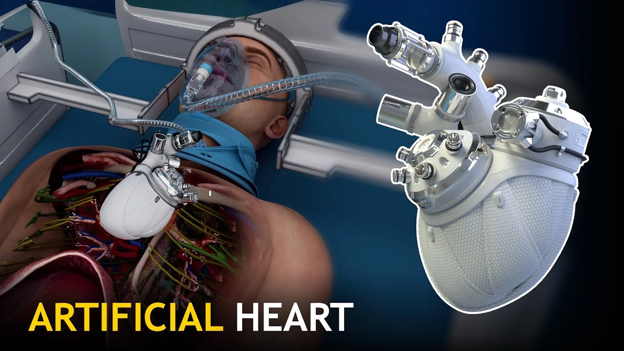
Artificial Heart: A Life-Saving Innovation in Modern Medicine – Medical marvels continue to advance rapidly, offering newfound hope for those facing critical health conditions. One of the most significant breakthroughs in cardiology is the development of the artificial heart. This device is far more than just an electronic gadget; it is a complex mechanical system designed to take over the vital role of the hardest-working organ in our body.
What Exactly is an Artificial Heart?
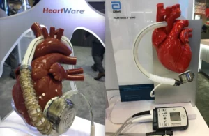
In simple terms, an artificial heart is a mechanical pump surgically installed to replace the function of the heart’s ventricles. In a healthy body, these two lower chambers are responsible for pumping blood to the lungs and throughout the entire system. When end-stage heart failure occurs and these chambers can no longer perform their duties, an artificial heart serves as a crucial medical intervention.
The installation of a Total Artificial Heart (TAH) involves the complete replacement of both lower chambers. The device is then connected directly to the upper chambers of the heart (the atria) and the main arteries. Through this connection, blood circulation that was previously obstructed can flow freely again, ensuring every tissue in the body receives an adequate oxygen supply.
A Vital Role as a “Bridge” to Life
It is essential to understand that, currently, an artificial heart is primarily used as a temporary solution. In the medical field, this is often referred to as a “Bridge to Transplant.” There are several reasons why this approach is taken:
-
Waiting for a Donor: Finding a compatible human heart donor is a lengthy process due to the limited number of available matches.
-
Maintaining Patient Stability: Patients with severe heart failure are often too weak to survive a long wait. The artificial heart maintains their health, ensuring they are strong enough when a donor heart finally becomes available.
-
Organ Recovery: By restoring healthy circulation, other organs—such as the kidneys and liver—that may have been struggling can recover before the major transplant surgery takes place.
How Does it Function?
While it may seem highly technical, the principle behind an artificial heart mimics the natural action of heart muscles. The device utilizes an external power source and a control system to regulate the pumping rhythm. Blood entering the atria is channeled into the mechanical sacs of the artificial heart and then pumped out with calibrated force to keep the patient’s blood pressure stable.
While using this device, patients are typically connected to an external driver (console) that monitors blood flow in real-time. Although this imposes certain physical limitations, the latest technology has allowed for more compact control units, enabling patients to maintain a degree of mobility within a hospital or home setting.
New Hope for Heart Failure Patients

Heart failure often leaves sufferers with severe shortness of breath, chronic fatigue, and swelling throughout the body. Intervening with an artificial heart can alleviate these heavy symptoms almost instantly once normal circulation is restored.
This technological leap proves that the line between biology and mechanical engineering is blurring for the sake of extending human life. For many families, the existence of the artificial heart is not just about surgical success rates—it is about gaining precious extra time with their loved ones.
The artificial heart stands as a milestone in modern medicine, providing a second chance for patients with permanent heart damage. As a temporary replacement for failed ventricles, this device ensures that life keeps beating while waiting for a permanent donor heart. Raising awareness about this technology is vital so that society understands that heart failure is no longer a definitive end, but a challenge that can be bridged with extraordinary innovation.
Medical Disclaimer: The decision to use an artificial heart is always based on a deep evaluation by a team of cardiac specialists. If you or a relative have concerns regarding heart function, consult a medical expert immediately for an accurate diagnosis.
New Era of Health: The Real-World Arrival of Wearable Kidneys
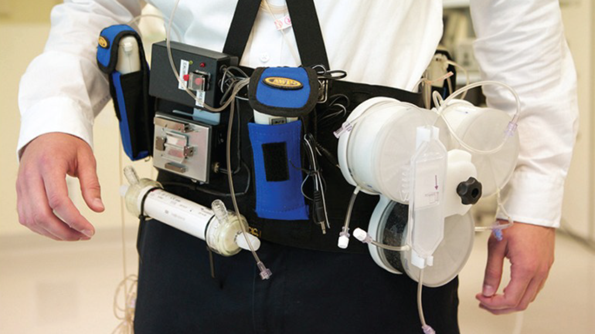
New Era of Health: The Real-World Arrival of Wearable Kidneys – Medical technology is constantly racing to bridge the gap between patient needs and the limitations of current equipment. For those living with kidney failure, daily routines often feel like a race against time, tethered to bulky machines in dialysis centers. However, a new beacon of hope is appearing on the medical horizon: the concept of the Wearable Artificial Kidney (WAK). This technology is more than just a medical aid; it is a promise of freedom for millions of patients worldwide.
Why Do We Need Portable Dialysis?

Traditionally, kidney failure patients who have not yet received a transplant rely heavily on conventional dialysis procedures. While this method is highly effective at filtering blood and removing excess fluid, the process takes hours and often leaves patients feeling profoundly exhausted. Furthermore, the physical confinement to a machine necessitates staying stationary, which automatically diminishes quality of life and productivity.
The quest to create a miniature dialysis machine actually began as far back as the 1970s. Unfortunately, technology at the time was unable to shrink machine components enough to make them light enough to carry. The main obstacles always revolved around battery weight, filter size, and the massive amount of pure water required for the dialysate fluid.
Technological Breakthroughs Behind Autonomous Devices
Now, thanks to intensive research in the United States and other developed nations, these technical hurdles are starting to clear. Scientists have successfully developed much lighter components and high-endurance batteries. The most crucial innovation lies in advanced filtration systems capable of recycling the dialysate fluid. This means the machine no longer requires gallons of pure water, allowing it to be sleek enough to be hidden under clothing.
Currently, development focuses on three primary types of devices:
-
Portable Hemodialysis Devices: Focused on direct blood cleaning.
-
Peritoneal Dialysis Devices: Utilizing the patient’s abdominal lining as a natural filter.
-
Hybrid Machines: Innovations that combine both methods for optimal results.
More Natural Health Benefits
The advantages of portable kidneys extend far beyond aesthetics or physical comfort. Medically, these devices mimic the function of a real kidney by filtering blood 24 hours a day. This continuous work cycle is much healthier than conventional dialysis, which is performed periodically (a few times a week).
With around-the-clock cleaning, a patient’s blood pressure tends to remain more stable. Additionally, fluid buildup in the body can be significantly minimized, which ultimately reduces the workload on the heart. Another piece of good news for patients is increased dietary flexibility; they no longer need to follow such restrictive diets because toxic waste is being constantly removed from their bloodstream.
WAK: From the Lab to Clinical Trials
One of the innovations showing the most tangible progress is the WAK (Wearable Artificial Kidney). This is currently the only device that has undergone human testing phases. In three initial studies, the results showed very promising performance.
The latest version of the WAK now weighs only about two pounds (less than one kilogram). This lightweight design is supported by a small battery that can be recharged while the patient sleeps at night. Although two more clinical study phases are required before it is ready for the general public, the existence of the WAK provides concrete proof that the future of portable dialysis is within reach.
Anticipating a Freer Future
The transition from giant machines to a device that can be worn like a belt certainly requires high precision. Researchers are committed to ensuring that every flaw found in previous trials is completely corrected before mass production begins. While it may take a few more years for these devices to become available at local hospitals, the progress happening every day is a strong reason to stay optimistic.
For those battling kidney failure, this technology is not just a medical tool—it is a ticket to returning to a normal life—working, traveling, and staying active without feeling “tied” to a life-sustaining machine. Let us keep a close eye on these developments, as this dream is one step closer to becoming a reality that will change the face of healthcare forever.
Bipolar vs. Monopolar: Choosing the Right Electrosurgery Mode
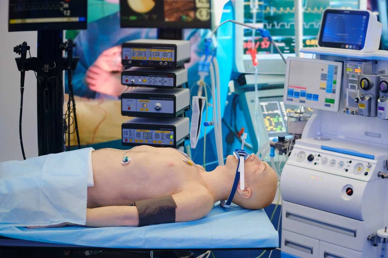
Bipolar vs. Monopolar: Choosing the Right Electrosurgery Mode – Modern surgical procedures have moved far beyond the traditional scalpel. In today’s operating rooms—particularly during high-precision tasks like ophthalmic surgery—surgeons rely heavily on a device known as the Electrosurgical Unit (ESU). This instrument has become indispensable due to its unique ability to cut tissue and control bleeding simultaneously using electrical energy.
The Physics of Electrical Energy in Tissue
The fundamental principle of electrosurgery lies in the application of high-frequency alternating current (AC). These frequencies typically range from 100 kilohertz to 5 megahertz, with voltages spanning from 200 to as high as 10,000 Volts. This current is passed directly through the tissue to generate controlled thermal energy.
The clinical objective of this heat varies depending on the specific surgical needs. A surgeon can adjust the device to cut through tissue, coagulate blood vessels, ablate specific growths, or even shrink tissue masses. This versatility is possible because the ESU generator can modify electrical waveforms. It is the change in these waveforms that dictates whether the tissue will be sliced cleanly or thickened (coagulated) to stop hemorrhaging.
Strategic Differences: Bipolar vs. Monopolar Methods

In clinical practice, electrosurgery is categorized into two primary modes, each with a distinct electrical circuit design. The choice between them depends entirely on the type of surgery and the required safety parameters.
1. Bipolar Electrosurgery: The “Wet Field” Specialist
The bipolar method is widely regarded as the safer and more precise option for delicate or confined spaces. In this system, both the active and return electrode functions are housed within a single instrument, usually shaped like a pair of forceps.
The electrical current only flows between the two tips of the forceps. This means only the specific tissue grasped by the instrument is part of the electrical circuit. A major advantage of this method is its ability to function in fluid-filled environments. This is why bipolar electrosurgery is the “gold standard” in eye or neurosurgery, as it can effectively coagulate even in a “wet field” where fluids are present.
2. Monopolar Electrosurgery: Versatility for Broader Areas
Unlike the bipolar system, the monopolar method places only the active electrode at the surgical site. To complete the electrical circuit and allow the current to flow, a patient return electrode (often called a “dispersive pad”) must be attached to another part of the patient’s body, such as the thigh or back.
The current travels from the surgical tip, through the patient’s body, and exits via the return pad. Because it covers a broader range, the monopolar system is highly efficient for surgeries involving larger tissue areas. However, this system requires meticulous attention to the placement of the return pad to prevent unintended risks.
Ensuring Patient Safety in the Operating Room
Advanced technology carries a high responsibility for safety. One of the most significant risks in monopolar ESU use is the potential for burns at the site of the return electrode. This occurs if the heat generated by the current is not safely dissipated or spread across a wide enough area.
To prevent this, the size and conductivity of the return pad must meet strict clinical standards. If the pad is not perfectly adhered or is too small for the patient, the heat will concentrate at a single point, potentially causing skin burns. Furthermore, the use of high frequencies (above 100 kHz) is intentional; it prevents the stimulation of the patient’s nerves and muscles, ensuring no involuntary muscle contractions or “shocks” occur during the procedure.
Electrosurgical Units have fundamentally changed how surgeons operate, offering a level of control far superior to traditional methods. By understanding the critical differences between precise bipolar techniques for wet environments and efficient monopolar techniques for broader applications, medical teams can ensure that procedures are not only fast but also meet the highest safety standards. For healthcare facilities, investing in high-quality ESU generators and maintaining a deep technical understanding of electrical waveforms is key to delivering optimal surgical care.

Robotic Surgery: Precision Beyond Human Hands
Robotic Surgery: Precision Beyond Human Hands – Medical technology is advancing at an incredible pace, pushing surgical precision to levels that were unimaginable just a few decades ago. One of the most significant leaps now becoming a standard in modern hospitals is robotic surgery. While the term “robotic” might spark images of autonomous machines, the reality in the operating room is far more collaborative and fascinating.
What is Robotic Surgery, Exactly?
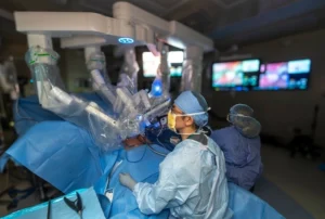
At its core, robotic surgery is a method where your surgeon utilizes a specialized robotic device to perform a procedure. This system typically consists of mechanical arms equipped with micro-surgical instruments designed for extreme maneuverability.
It is crucial to emphasize one point: the robot does not replace the surgeon. These machines do not make independent decisions or perform incisions on their own. Instead, the surgeon remains in total control throughout the process. Sitting at a high-tech console featuring a high-definition 3D display, the surgeon moves the robotic arms using sensitive controllers. Every hand movement is translated in real-time into precise, scaled-down motions of the instruments inside the patient’s body.
Why Precision is the Game Changer
In traditional surgery or even standard laparoscopy, a surgeon’s range of motion can sometimes be limited by human anatomy or natural, involuntary hand tremors. This is where robotic-assisted technology shines.
-
Tremor Filtration: The robotic system is designed to filter out microscopic hand tremors, ensuring that instrument movements are incredibly steady and smooth.
-
Enhanced Range of Motion: Robotic arms possess “wristed” flexibility that exceeds the capabilities of the human hand. This allows instruments to rotate and reach tight corners within the body with pinpoint accuracy.
-
Superior 3D Visualization: The surgeon’s display provides a highly detailed, magnified 3D view. This allows for the clear identification of tiny nerves and blood vessels, significantly reducing the risk of accidental damage to surrounding tissues.
Tangible Benefits for the Patient
For patients recommended for this procedure, the primary advantages are often felt during the recovery phase. Because the instruments are so delicate and precise, the incisions required are much smaller than those in traditional open surgery—a hallmark of minimally invasive surgery.
This leads to a positive domino effect for patient health:
-
Reduced Pain: Smaller incisions mean less trauma to the body’s tissues, significantly lowering post-operative discomfort.
-
Lower Risk of Infection: Smaller wounds close faster and are less susceptible to external complications.
-
Faster Recovery Times: Patients typically require shorter hospital stays and can return to their daily routines much sooner than with conventional methods.
-
Minimal Blood Loss: The precision of the robot helps surgeons control blood vessels more effectively, reducing the need for blood transfusions.
Safety and the Surgeon’s Expertise

Understanding that this technology is an “advanced tool” in the hands of an expert is vital. Robotic surgery is an extension of a surgeon’s skill, experience, and clinical judgment. Before ever touching a robotic console, surgeons undergo rigorous specialized training and certification to ensure they can navigate the system with absolute safety.
From Pakistan to the global stage, the adoption of robotics in urology, gynecology, and even cardiac surgery is rising rapidly. It proves that the integration of human intuition and mechanical precision is the current reality of modern healthcare.
Robotic surgery is no longer just a futuristic trend; it is a practical solution for improving clinical outcomes. By offering unmatched precision, enhanced visualization, and an efficient recovery process, this method helps healthcare providers deliver safer and more effective care. If you or a loved one are considering a surgical procedure, discussing the option of robotic-assisted surgery with your medical team could be a smart step toward optimal healing.
AED: The Critical Bridge in Cardiac Survival
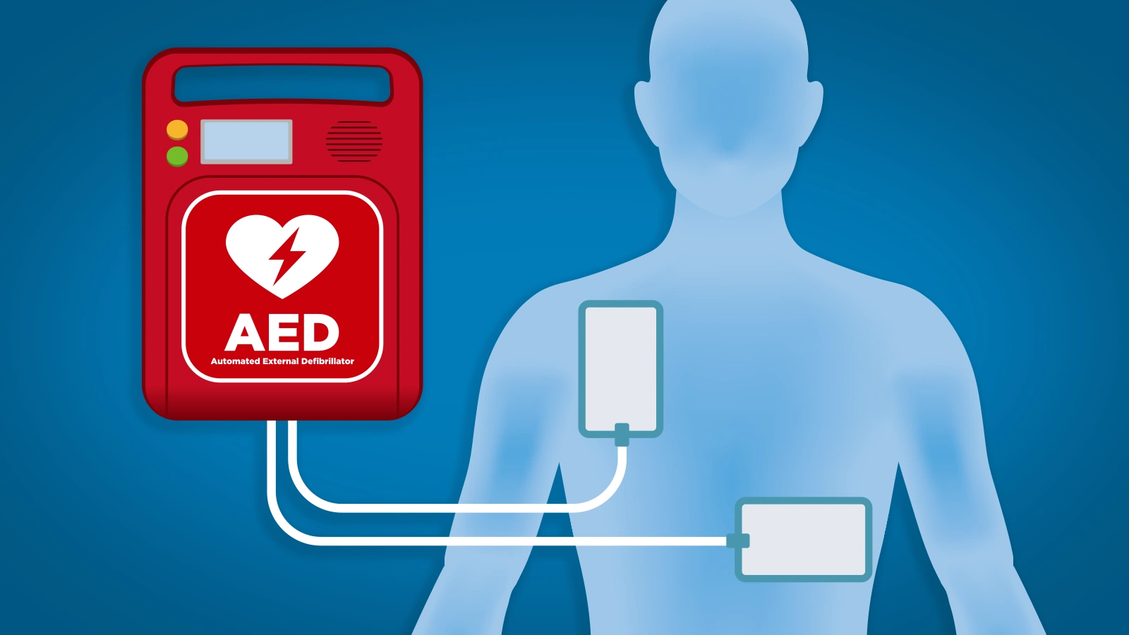
AED: The Critical Bridge in Cardiac Survival – Sudden cardiac death often strikes without warning. In emergency situations such as Sudden Cardiac Arrest (SCA), every second is a battle against time. This is where the Automated External Defibrillator (AED) plays a pivotal role. This portable medical device is engineered to be the first line of defense in saving lives before professional medical help arrives on the scene.
Currently, AEDs are becoming increasingly common in public spaces, ranging from airports and shopping malls to high-rise office buildings. However, public understanding of when and how these devices function must be improved to ensure their life-saving potential is fully realized.
Understanding Sudden Cardiac Arrest
Sudden cardiac arrest occurs when the heart’s electrical system malfunctions. This causes the heart to beat irregularly and dangerously fast (ventricular fibrillation), leading to a total failure in pumping blood to the rest of the body. Without blood flow, the brain and other vital organs are deprived of oxygen within seconds.
Permanent brain damage can occur in just a matter of minutes if the heart rhythm is not immediately restored to normal. While Cardiopulmonary Resuscitation (CPR) is vital for maintaining temporary blood flow, it is often only a shock from a defibrillator that can “reset” the heart’s rhythm back to stability. The synergy between CPR and the use of an AED has been clinically proven to drastically increase a patient’s chances of survival.
Do You Need an AED at Home?
While AEDs are widely available in public, a common question arises: Is it necessary to have one at home? The answer depends largely on the health profile of the household members.
For individuals with a history of severe coronary artery disease, chronic heart rhythm disorders, or those who have previously suffered a heart attack, having an AED at home can be a wise safety investment. Modern versions for home use are lightweight, easy to operate, and available in many regions without a prescription. However, it is highly recommended to consult with a cardiologist before making a personal purchase.
Advanced, User-Friendly Technology
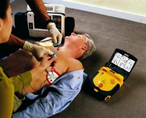
One of the primary advantages of modern AEDs is their ease of operation. Even if you are not a trained medical professional, you can still use this device effectively during an emergency.
In general, here is how an AED functions:
-
Automated Voice Instructions: Once activated, the AED provides step-by-step voice prompts. It will guide you on when to attach the electrode pads and when to stay clear of the patient.
-
Autonomous Analysis: These devices are highly intelligent. The AED automatically analyzes the patient’s heart rhythm. If the machine detects that a shock is not needed, it will not deliver one, making it incredibly safe to use without the risk of accidental harm.
-
Portable Design: With a compact build, the device is easy to carry and store, making it the new gold standard in modern emergency safety equipment.
Why Training Still Matters
Although AEDs are designed for anyone to use, undergoing Basic Life Support (BLS) training is highly recommended. Such training helps an individual remain calm under pressure and understand the proper coordination between performing chest compressions (CPR) and operating the AED machine. The courage to act and the speed of response are the ultimate keys to the cardiovascular chain of survival.
Healthcare technology like the AED has brought emergency room capabilities into the public domain. This device is more than just an electronic tool; it is the bridge between life and death for someone experiencing cardiac arrest. Understanding how an AED works and knowing its location in our surroundings is a small step that can have a massive impact on human life.
Sight Pakistan Insights: As a global provider of medical technology insights, we view the increasing adoption of AEDs in public facilities as a progressive step in public health. Ensure your medical devices are always “rescue-ready” by performing regular battery and electrode pad checks.

AI-ECG Wearables: Real-Time Cardiac Monitoring Trends
AI-ECG Wearables: Real-Time Cardiac Monitoring Trends – The Electrocardiogram (ECG) has been the cornerstone of cardiac diagnostics for over a century. However, in this digital era of 2026, this conventional tool is undergoing a profound “rebirth” driven by Artificial Intelligence (AI). The synergy between machine learning and ECG is not merely a software update; it represents a transformative shift in how clinicians interpret the human heartbeat.
Integrating AI into ECG interpretation allows for the detection of subtle electrical patterns that are often invisible to the human eye. While traditional ECGs were primarily used to assess a patient’s current cardiac state, AI-enhanced technology is pushing the boundaries into highly accurate predictive analytics.

Beyond Human Vision: The Edge of AI-ECG
The most compelling breakthrough in AI-enhanced ECG technology lies in its ability to identify structural heart diseases through electrical signals that appear perfectly normal to even the most seasoned cardiologists. Sophisticated algorithms, trained on millions of patient data points, can recognize minute anomalies that serve as early indicators of heart failure or valvular disorders.
Key pillars of this modern renaissance include:
-
Enhanced Diagnostic Accuracy: AI processes data objectively, significantly reducing the risk of interpretive errors caused by human fatigue or subjective bias.
-
Predictive Cardiovascular Events: This technology can now forecast the risk of future Atrial Fibrillation (AFib) or strokes, often before any clinical symptoms manifest in the patient.
-
Continuous Monitoring via Wearables: Thanks to AI, ECG features on smartwatches now possess serious clinical capabilities. Monitoring is no longer confined to hospital walls; it occurs in real-time, 24/7, during a patient’s daily activities.
The Challenges Behind the Algorithm
Despite the massive potential of AI-ECG, its journey toward mass implementation in global healthcare still faces significant hurdles. Medical technology is not just about digital prowess; it is deeply rooted in ethics and trust.
A primary challenge for health-tech developers today is the quality of training data. An AI algorithm is only as accurate as the data it consumes. If the datasets used are not diverse—spanning various ethnicities, ages, and genders—biases may emerge that could jeopardize patient safety.
Furthermore, legal accountability and explainability remain under the spotlight. Clinicians need to understand why an AI arrived at a specific diagnosis. Without a logical “paper trail” for the decision, it is difficult for medical professionals to rely entirely on a machine’s output. There are also valid concerns regarding the potential “de-skilling” of doctors if they become overly reliant on automation.
Shaping the Future of Cardiovascular Management
The presence of AI in the world of ECG is intended to serve as an intelligent assistant that augments the doctor’s role, not replaces it. With higher precision, patient care can become truly personalized. Each individual receives treatment tailored to a unique risk profile decoded from their specific ECG data.
In the coming years, we will witness a healthcare ecosystem where heart disease diagnosis is no longer reactive (treating after the fact) but proactive. Early detection fueled by AI-ECG will slash global healthcare costs and, most importantly, save countless lives through timely intervention.
The modern resurgence of the ECG through AI is proof that classic technology can remain relevant if it adapts. By prioritizing ethical standards, rigorous clinical validation, and robust patient data protection, the integration of AI-ECG will become the new gold standard in cardiovascular medicine. The ultimate focus remains unchanged: safeguarding the human heart with sharper intelligence and broader reach.
ECG Basics: Understanding Heart Electrical Activity
ECG Basics: Understanding Heart Electrical Activity – Heart health is often likened to a high-precision engine driving every bodily function. To ensure this “engine” operates smoothly, medical professionals rely on sophisticated technology capable of reading the subtle, rhythmic signals generated by every beat. Among the most essential instruments in cardiology is the Electrocardiogram, or ECG. Through a series of waves that may seem complex at first glance, an ECG tells a profound story about how electricity flows through the heart muscle to keep blood circulating effectively.
What Is an ECG and Why Is It Vital?
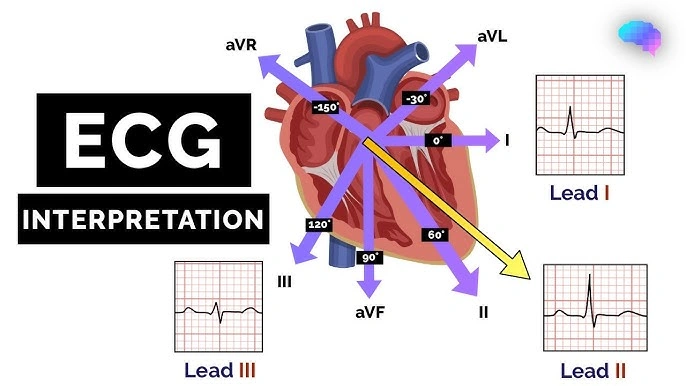
At its core, an ECG is a diagnostic procedure used to record the heart’s electrical activity from multiple angles. Its primary purpose is to detect pathologies and abnormalities in cardiac function. During a standard procedure, medical staff place electrodes on specific points across the patient’s limbs and chest. These sensitive sensors capture the electrical impulses that trigger heart contractions, visualizing them as waveforms on a monitor or printout.
The standard 12-lead ECG is widely considered the gold standard in diagnostics. This recording allows physicians to observe a “normal sinus rhythm”—a state where electrical signals travel through the heart’s natural pathways at a steady, regulated speed. However, beneath every single heartbeat lies a series of distinct components, each carrying critical medical significance.
Decoding ECG Waveforms: Breaking Down the Components
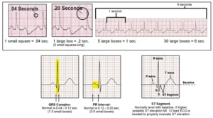
To grasp how an ECG functions, one must understand what each peak and valley in the graph represents. Every movement corresponds to a specific phase in the cardiac cycle:
-
The P Wave: The Starting Signal The P wave represents atrial depolarization. In healthy individuals, every heartbeat should be preceded by a P wave. If this wave is absent or irregular, it serves as an early warning sign of potential issues in the upper chambers of the heart.
-
The PR Interval: The Communication Bridge Spanning from the onset of the P wave to the beginning of the Q wave, the PR interval measures the time taken for electrical activity to travel from the atria to the ventricles. This brief pause is essential; if the interval is too long, it may indicate a blockage or delay in the heart’s electrical conduction system.
-
The QRS Complex: The Powerhouse This is the most prominent feature on an ECG graph. Comprised of three closely linked waves (Q, R, and S), the QRS complex represents ventricular depolarization. This phase generates the primary force required to pump blood to the entire body.
-
The ST Segment: The Contraction Phase Located between the end of the S wave and the start of the T wave, the ST segment is an isoelectric line representing the period between ventricular depolarization and repolarization—essentially, the time when the ventricles are contracting to eject blood. Shifts in this line (elevations or depressions) are often critical indicators of acute cardiac events, such as a heart attack.
-
The T Wave: The Recovery Phase After pumping, the heart must “recharge” its energy. The T wave represents ventricular repolarization, or the recovery phase. Although these waves appear small, abnormalities here can provide clues regarding electrolyte imbalances or chronic blood flow issues.
RR and QT Intervals: Measuring Speed and Duration
Beyond waveform shapes, clinicians pay close attention to the time elapsed between beats. The RR interval, measured from the peak of one R wave to the next, is used to calculate the heart rate per minute. Meanwhile, the QT interval encompasses the entire process from the start of ventricular contraction to the end of recovery. Monitoring the QT interval is vital, as an excessively long duration can increase the risk of dangerous heart rhythm disorders.
Why Expert Interpretation Matters
While the basic principles of an ECG seem straightforward, interpreting real-world results can be highly complex. Waveform variations across patients can be vast, influenced by age, underlying conditions, and lifestyle factors. Consequently, medical practitioners often refine their diagnostic skills by studying large clinical case banks, analyzing hundreds of real ECGs to understand the nuances of step-by-step interpretation.
Mastering ECG analysis is about more than just reading moving lines; it is about understanding the rhythm of life itself. With accurate technology and sharp interpretive skills, healthcare providers can save lives by detecting heart disorders long before they escalate into life-threatening emergencies.
Modern Pacemakers: Technology and Clinical Benefits
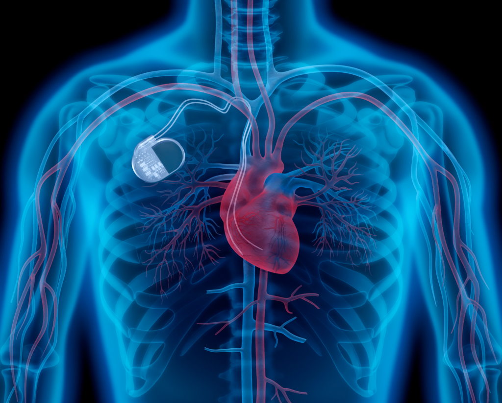
Modern Pacemakers: Technology and Clinical Benefits – Managing a life-threatening heart rhythm disorder often requires advanced medical intervention. For many patients, doctors recommend the implantation of a pacemaker—a sophisticated, life-saving device designed to keep the heart beating in a consistent, healthy rhythm. If you or a loved one are facing cardiac rhythm challenges, understanding how this technology works and why it is used is the first step toward effective treatment.
What Is a Pacemaker and How Does It Function?
At its core, a pacemaker is a small, battery-operated electronic device. Despite its compact size, it performs a crucial role: monitoring the heart’s electrical activity and sending low-energy electrical impulses when necessary. These impulses encourage the heart to beat at a steady, normal rate, ensuring that the heart is neither too slow nor excessively fast. By maintaining this regulated rhythm, the device allows the heart to pump blood effectively to the rest of the body, preventing complications such as fatigue, fainting, or more severe cardiovascular events.
When Is a Pacemaker Necessary?
Medical professionals typically suggest a pacemaker for individuals suffering from arrhythmias, a condition where the heart’s natural electrical system malfunctions. While arrhythmia is the most common reason for implantation, these devices are also critical tools in the management of specific cases of heart failure and post-heart attack recovery. In these scenarios, the pacemaker acts as a safeguard, stepping in to ensure that the heart’s workload remains manageable and that the body receives adequate oxygenated blood flow.
Critical Assessments Prior to Implantation
Deciding to undergo a procedure to install a pacemaker is a significant medical step. Consequently, healthcare providers perform a comprehensive series of diagnostic tests to ensure that the heart truly requires external electrical support. Every patient is unique, and these assessments provide the clinical data necessary to tailor the device’s settings to the individual’s specific needs.
Standard diagnostic protocols before the procedure often include:
-
General Physical Examination: A holistic assessment of overall health to determine surgical eligibility and baseline cardiovascular status.
-
Electrocardiogram (ECG/EKG): A standard test that records the electrical signals in the heart, providing a clear snapshot of current rhythm abnormalities.
-
Holter Monitoring: Unlike a standard ECG, this portable device records the heart’s activity over 24 to 48 hours, capturing intermittent issues that might not appear during a brief clinical visit.
-
Echocardiogram: This ultrasound-based imaging test provides a visual look at the heart’s structure, allowing doctors to evaluate how well the heart valves and chambers are pumping.
-
Stress Test: Often performed while walking on a treadmill or using a stationary bike, this test measures how the heart performs under physical exertion, helping identify limitations in heart rhythm response.
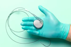
Living with the Device
Modern advancements have made pacemakers smaller and more durable than ever before. While the adjustment period after surgery varies from person to person, most patients report a significant improvement in their quality of life. The device essentially operates in the background, allowing individuals to return to their daily activities with the peace of mind that their heart rhythm is being monitored and corrected in real-time.
Advancements in medical technology continue to bridge the gap between complex heart conditions and a normal, active lifestyle. A pacemaker is far more than just an electrical gadget; it is a vital component of modern cardiac care that bridges the gap between potential danger and a healthy heart rate. If you have been advised to undergo this procedure, rest assured that these diagnostic steps and the device itself are proven methods to stabilize your cardiovascular health.
Always maintain open communication with your cardiologist regarding your symptoms and lifestyle needs, as they are the best resource for managing your specific heart health journey.