How AeviceMD Is Advancing Patient Monitoring and Cardiovascular Care
How AeviceMD Is Advancing Patient Monitoring and Cardiovascular Care | Healthcare is evolving rapidly as hospitals and medical professionals adopt smarter technologies to improve patient outcomes. Digital health solutions, wearable medical devices, and connected monitoring systems are transforming the way clinicians diagnose, monitor, and manage diseases. Instead of relying solely on periodic hospital visits, healthcare providers can now access continuous patient data that supports faster interventions and more personalized treatment plans.
Among the innovators driving this transformation is AeviceMD, a medical technology company focused on wearable patient monitoring solutions. By combining advanced sensor technology with digital healthcare platforms, AeviceMD aims to help healthcare professionals monitor patients more effectively while improving comfort and convenience for users.
Beyond wearable monitoring, modern healthcare also depends on sophisticated diagnostic technologies such as ultrasound imaging, pacemakers, and other cardiovascular devices. Together, these innovations are helping reshape patient care by making healthcare more proactive, connected, and data-driven.
This article explores how AeviceMD contributes to digital healthcare, the importance of continuous patient monitoring, and why cardiovascular technologies remain essential in modern medicine.
The Growing Need for Continuous Patient Monitoring
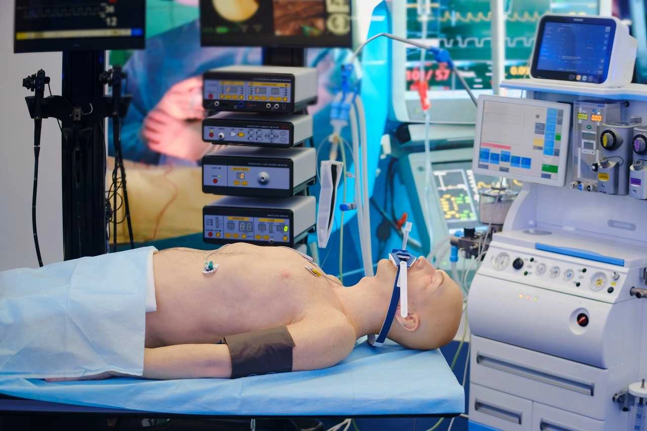
Modern medicine increasingly emphasizes prevention and early intervention rather than simply treating illnesses after symptoms become severe.
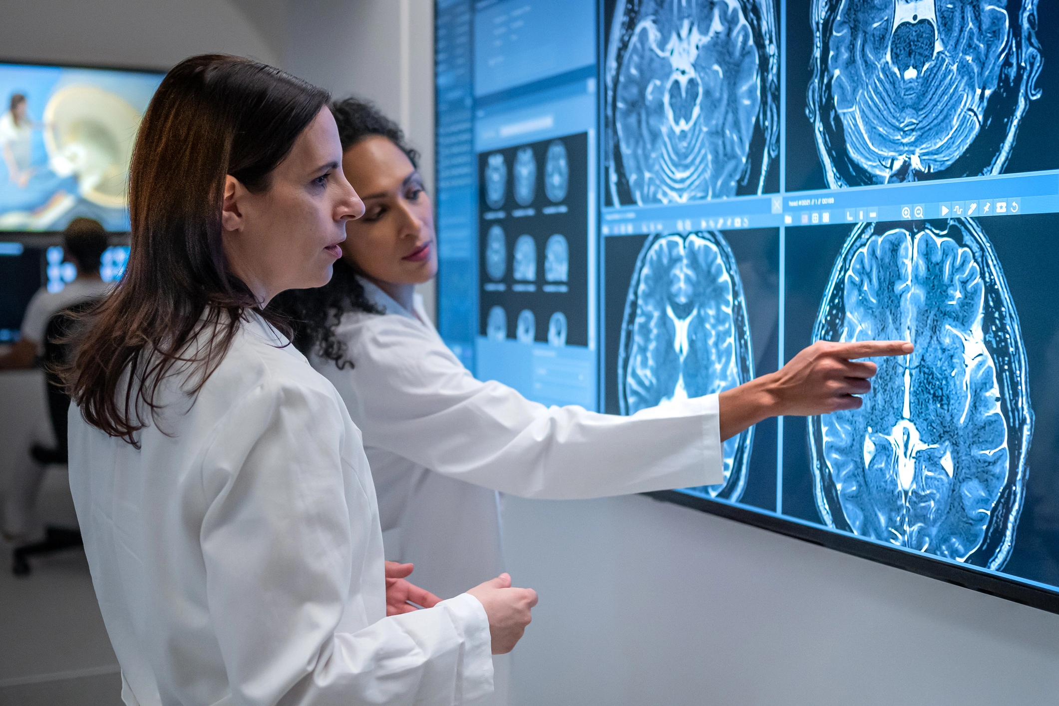
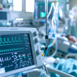
Continuous patient monitoring allows healthcare professionals to observe changes in vital signs over extended periods, providing valuable clinical insights that may not be captured during routine hospital visits.
This approach is especially important for individuals living with chronic respiratory diseases, cardiovascular conditions, and other long-term health issues that require ongoing observation.
By detecting subtle physiological changes early, healthcare providers can make faster decisions, adjust treatment plans, and potentially prevent serious medical complications.
Key Insight: Continuous monitoring enables earlier detection of health changes, helping clinicians deliver more timely and personalized care.
AeviceMD’s Mission in Digital Healthcare
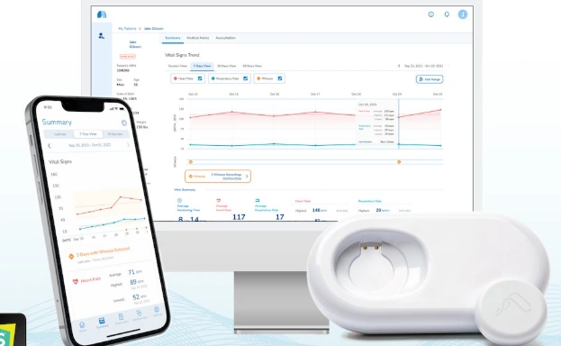
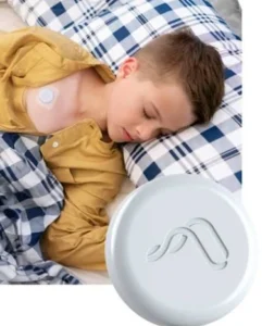
AeviceMD focuses on developing wearable medical technologies designed to support remote patient monitoring and continuous health assessment.
Its solutions are intended to bridge the gap between hospital-based care and everyday life by allowing patients to be monitored outside traditional clinical environments.
Rather than replacing healthcare professionals, wearable technologies provide additional clinical information that supports more informed medical decision-making.
As healthcare systems increasingly embrace digital transformation, companies like AeviceMD are helping improve accessibility, efficiency, and patient engagement through innovative monitoring solutions.
Why Wearable Medical Devices Matter
Wearable healthcare technology has become one of the fastest-growing areas of medical innovation.
Unlike traditional monitoring systems that require patients to remain in hospitals, wearable devices enable continuous observation while individuals continue their normal daily activities.
Some of the major benefits include:
- Continuous monitoring of physiological data.
- Greater patient mobility and comfort.
- Earlier detection of health changes.
- Improved long-term disease management.
- Reduced need for frequent hospital visits.
- Better communication between patients and healthcare providers.
These advantages support more proactive healthcare while improving overall patient experience.
The Role of Patient Monitoring in Chronic Disease Management
Millions of people worldwide live with chronic illnesses that require ongoing observation.
Patients with respiratory disorders, cardiovascular diseases, hypertension, or chronic obstructive pulmonary disease (COPD) often benefit from continuous monitoring that helps healthcare providers track disease progression over time.
Instead of relying only on occasional appointments, physicians can evaluate long-term health trends, making treatment decisions based on more comprehensive information.
This data-driven approach supports personalized healthcare and may improve long-term patient outcomes.
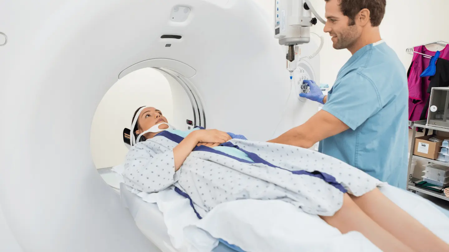
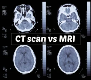
Ultrasound Technology in Modern Medicine
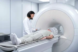
Ultrasound imaging remains one of the most important diagnostic tools available to healthcare professionals.
Using high-frequency sound waves, ultrasound systems produce real-time images of internal organs, muscles, blood vessels, and other body structures without exposing patients to ionizing radiation.
Healthcare providers commonly use ultrasound for:
- Cardiology
- Obstetrics
- Emergency medicine
- Internal medicine
- Vascular examinations
- Musculoskeletal assessments
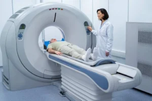
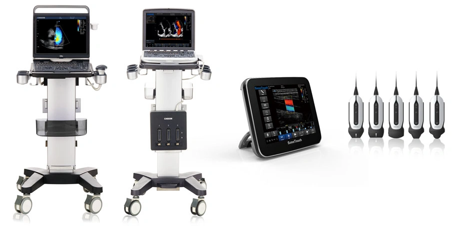
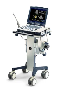
Advances in portable ultrasound technology have also expanded access to diagnostic imaging in outpatient clinics, rural healthcare settings, and emergency care environments.
Cardiovascular Devices That Improve Patient Care
Heart disease continues to be one of the leading health challenges worldwide, making cardiovascular technology an essential part of modern medicine.
Medical devices used in cardiology help physicians diagnose, monitor, and treat a wide range of heart conditions while improving patient safety and quality of life.
Common cardiovascular technologies include:
- Pacemakers
- Implantable cardioverter-defibrillators (ICDs)
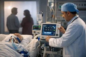
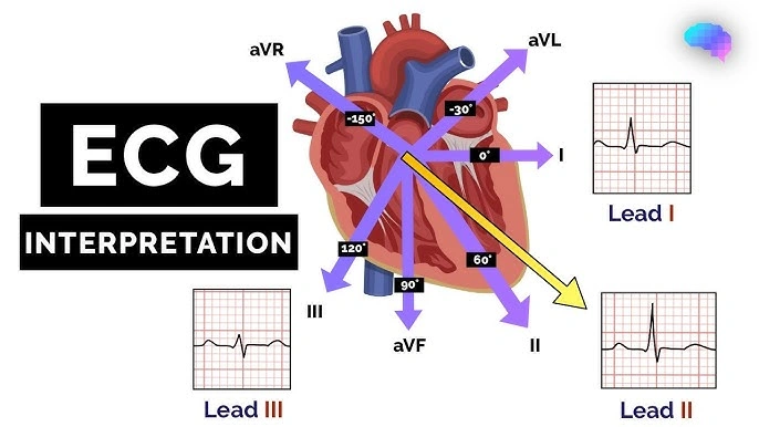
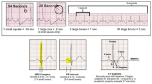
- Electrocardiogram (ECG) systems
- Cardiac monitoring devices
- Holter monitors
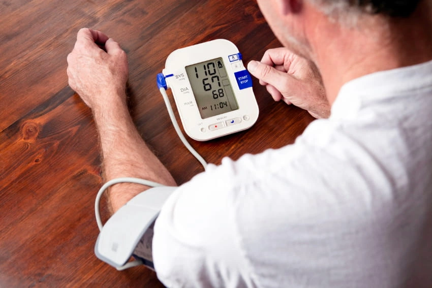
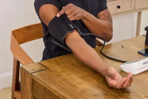
- Blood pressure monitoring equipment
Each technology plays a unique role in supporting cardiovascular health throughout diagnosis, treatment, and long-term management.
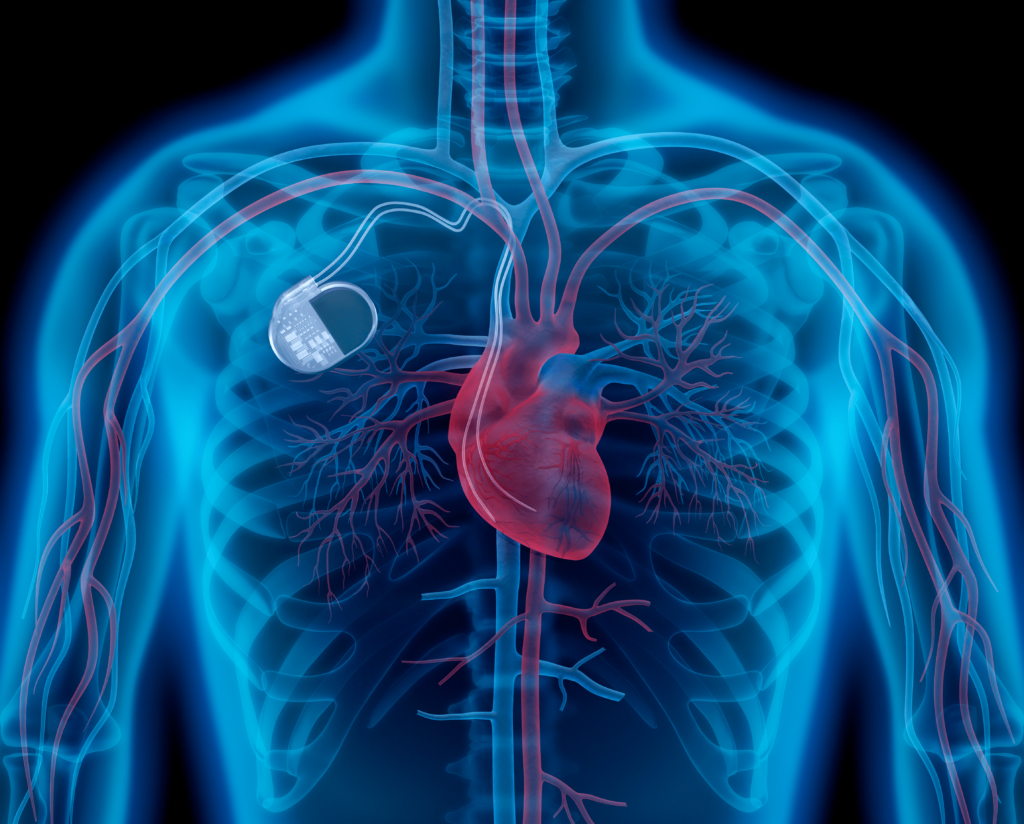
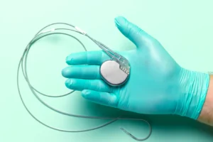
Understanding Pacemakers
Pacemakers are implantable medical devices designed to regulate abnormal heart rhythms.
When the heart beats too slowly or irregularly, the pacemaker delivers controlled electrical impulses that help maintain a stable heartbeat.
Modern pacemakers have evolved significantly over the years, offering improved reliability, longer battery life, and advanced monitoring capabilities.
For many patients with cardiac rhythm disorders, these devices improve daily life while reducing the risk of serious complications.
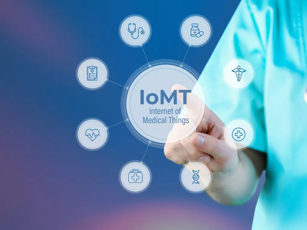
Digital Health Is Transforming Cardiology
Cardiovascular medicine has become one of the leading beneficiaries of digital health innovation.
Remote monitoring systems, wearable sensors, cloud-connected healthcare platforms, and artificial intelligence are enabling clinicians to monitor patients more efficiently than ever before.
These technologies support:
- Earlier diagnosis.
- Continuous patient observation.
- Personalized treatment planning.
- Reduced hospital admissions.
- Better long-term disease management.
- More efficient healthcare delivery.
As digital healthcare continues to evolve, connected technologies are expected to become increasingly integrated into cardiovascular care.
Benefits for Healthcare Providers
Advanced monitoring technologies offer significant advantages not only for patients but also for healthcare professionals.
Clinicians benefit from access to larger amounts of clinical data, allowing them to identify health trends that might otherwise go unnoticed.
These technologies help healthcare providers:
- Improve diagnostic accuracy.
- Monitor patients remotely.
- Prioritize high-risk cases.
- Support evidence-based clinical decisions.
- Enhance workflow efficiency.
- Improve long-term patient follow-up.
Together, these improvements contribute to higher-quality healthcare services.
The Future of Connected Healthcare
Healthcare is becoming increasingly connected through digital innovation.
Future developments are expected to include:
- Artificial intelligence-assisted diagnostics.
- Expanded remote patient monitoring.
- Predictive health analytics.
- Personalized treatment recommendations.
- Greater integration between wearable devices and hospital information systems.
- Increased use of cloud-based healthcare platforms.
Companies developing innovative patient monitoring technologies today are helping shape a future where healthcare becomes more accessible, efficient, and personalized.
Frequently Asked Questions
What is AeviceMD?
AeviceMD is a medical technology company that develops wearable patient monitoring solutions designed to support continuous health assessment and remote clinical care.
What is patient monitoring?
Patient monitoring involves continuously collecting physiological information to help healthcare professionals evaluate health conditions and detect changes that may require medical attention.
Why are wearable medical devices important?
Wearable devices enable continuous health monitoring outside hospitals, supporting earlier diagnosis, better disease management, and greater patient convenience.
What are ultrasound systems used for?
Ultrasound systems use sound waves to produce real-time images of internal organs, blood vessels, muscles, and other body structures for medical diagnosis.
What does a pacemaker do?
A pacemaker regulates abnormal heart rhythms by sending electrical impulses that help maintain a stable heartbeat.
The future of healthcare depends on technologies that improve access to medical information while supporting better patient outcomes. AeviceMD represents this new generation of healthcare innovation by developing wearable patient monitoring solutions that help bridge the gap between hospitals and everyday life.
Together with established technologies such as ultrasound systems, pacemakers, and other cardiovascular devices, continuous patient monitoring is helping transform medicine into a more proactive, data-driven, and patient-centered field. As digital health continues to advance, these innovations will play an increasingly important role in improving diagnosis, treatment, and long-term disease management across healthcare systems worldwide.