Next-Generation Sequencing for Modern Pathology
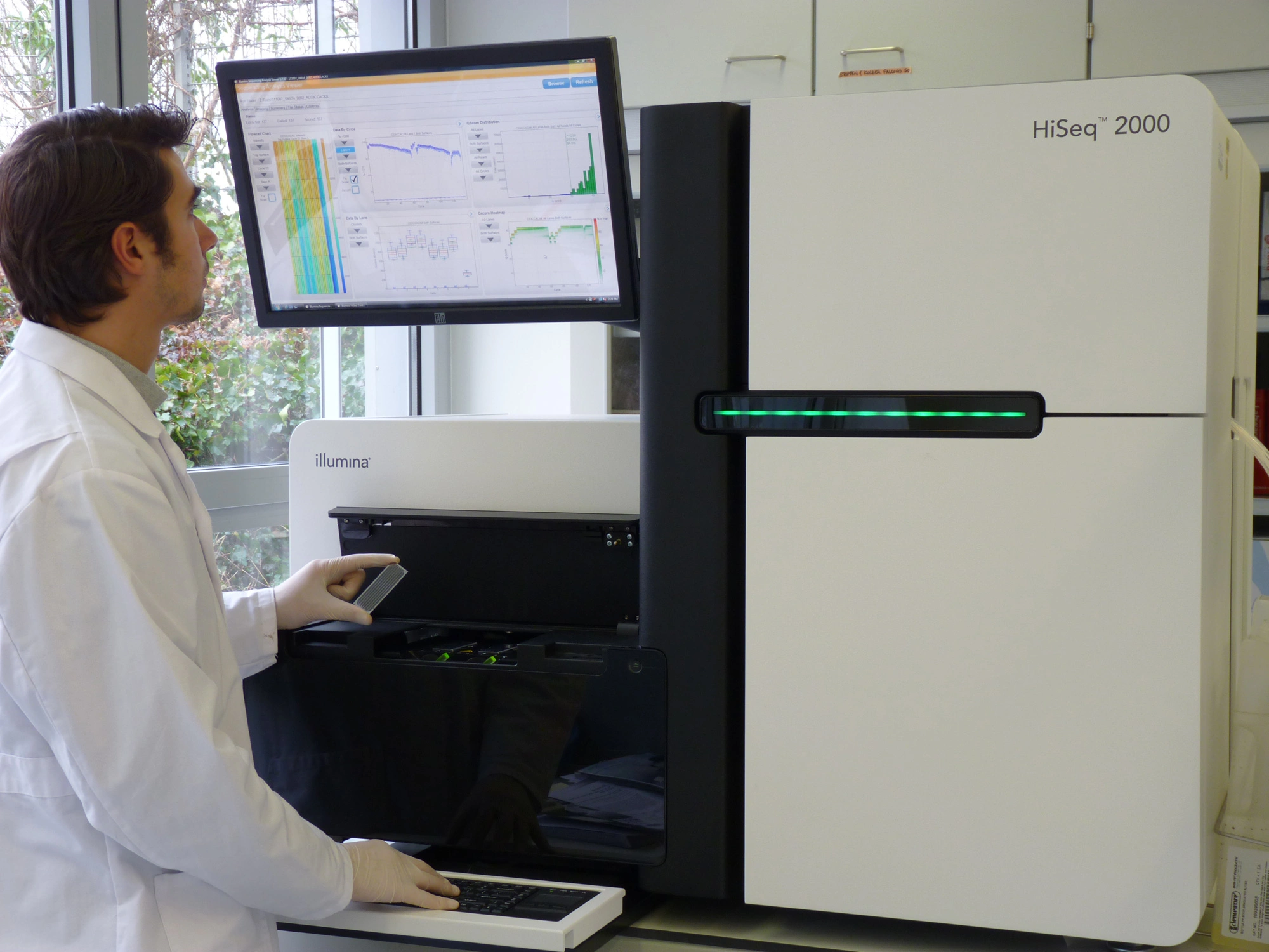
Next-Generation Sequencing for Modern Pathology | Modern medical diagnosis no longer relies solely on observing physical symptoms or conventional microscopic examinations. The emergence of large-scale DNA and RNA analysis technologies has fundamentally changed how medical professionals understand diseases. At the forefront of this transformation is a method known as Next-Generation Sequencing (NGS).
This technology works by reading millions of nucleic acid strand fragments simultaneously. This capability significantly shortens the process of mapping the human genetic blueprint, making it highly efficient while covering a vast area in a single test. For pathologists and clinical laboratory managers, this innovation opens the door to far more precise and personalized diagnostic determinations.
Bridging Translational Research and Clinical Diagnostics

Today’s laboratories are frequently faced with two major responsibilities at once. On one hand, they must support translational research to discover new biomarkers. On the other hand, they are required to provide rapid routine diagnostic services for patient safety. Answering this dual challenge, Thermo Fisher Scientific designed an NGS ecosystem that is adaptive and functionally integrated.
The software and instruments developed within this line ensure that genetic testing workflows run in alignment with medical regulatory standards. Through a comprehensive approach, researchers can easily transition laboratory findings into real-world clinical applications. This cuts through the technical red tape that usually hinders integration between the research community and frontline healthcare services.
Core Pillars of Modern NGS Technology Excellence
Implementing sequencing technology in healthcare facilities demands exceptionally strict standards. Various structural advantages are intentionally embedded into this modern NGS system to ensure daily operational effectiveness in the laboratory:
High Accuracy on a Macro Scale
Misreading a single nitrogenous base can be fatal for clinical interpretation, especially when detecting rare mutations in cancer. This sequencing system utilizes advanced chemistry capable of identifying genetic variants with remarkably high sensitivity, minimizing the risk of false-negative results.
Rapid Turnaround Time
When managing critical patients or determining advanced-stage cancer therapies, time is an invaluable asset. The latest NGS platforms can slash waiting times from weeks to days, or even hours. This speed allows specialists to make therapeutic decisions immediately without delaying patient care.
Simple Workflow Automation
One of the biggest hurdles in adopting conventional NGS is the complexity of sample preparation, or library preparation. Through end-to-end automation, manual operator involvement (hands-on time) is drastically reduced. Consequently, the potential for human error can be minimized.
Operational Cost Efficiency
The assumption that genetic mapping tests are always capital-intensive is now beginning to fade. The efficient use of reagents and maximization of sequencing chip capacity make the cost per sample far more economical. This cost structure helps hospitals manage their operational budgets more wisely.
Practical Transformation in the Pathology Room

The presence of a compact NGS ecosystem brings a tangible impact to the daily activities of pathologists. When analyzing tumor biopsy tissue where sample volume is extremely limited, older sequencing methods often deplete the tissue material before all target genes can be examined.
Utilizing targeted NGS panels allows for the simultaneous examination of tens to hundreds of relevant genes from just a single, small biopsy specimen. Pathologists can identify point mutations, insertions, deletions, and gene fusions in great depth. This comprehensive genomic information serves as the core foundation for generating accurate molecular pathology reports.
Supporting the Implementation of Precision Medicine
Every patient possesses a unique genetic profile, which means their responses to medications vary. This is where personalized medicine, or precision medicine, plays a vital role. The genomic data generated by NGS instruments provides a thorough overview of a disease’s biological vulnerabilities.
For example, in oncology management, sequencing results can indicate whether a patient will respond to a specific targeted therapy or display resistance instead. Furthermore, in pharmacogenomics, this data helps predict how a patient’s body metabolizes drugs, enabling physicians to determine optimal dosages and avoid life-threatening side effects.
Automation Eliminates Technical Hurdles in the Lab
For years, sequencing technology was considered the exclusive domain of large-scale research laboratories that employed dedicated bioinformatics experts. The complexity of raw data analysis was often a major deterrent for mid-sized healthcare facilities.
To overcome this constraint, Thermo Fisher Scientific’s NGS platforms come equipped with integrated data analysis systems that translate raw sequencing data into easy-to-read clinical reports. Laboratory personnel no longer need to perform tedious manual data processing. Built-in artificial intelligence and databases automatically match discovered genetic variants with the latest medical literature, generating clinical recommendations ready for medical team review.
Looking to the Future of Molecular Diagnostics
The need for genetic testing is projected to rise sharply alongside growing awareness of early disease detection. Choosing the right NGS infrastructure is not just a short-term medical equipment investment; it is a strategic long-term step to elevate an institution’s healthcare capabilities.
By providing reliable, flexible tools backed by a robust ecosystem, laboratories can continue to thrive alongside the current of medical innovation. Mastering genomic technology ultimately converges on one main objective: saving more lives through faster, more accurate, and dependable diagnoses for the wider community.
Essential NICU Equipment for Neonatal Care
Essential NICU Equipment for Neonatal Care | The birth of a child is naturally a moment filled with profound joy. However, for some parents, this experience can quickly shift into a phase of intense anxiety when their newborn arrives prematurely or faces critical health complications. These vulnerable infants require highly specific, intensive medical intervention within the Neonatal Intensive Care Unit (NICU).
The NICU is far from an ordinary hospital ward. This specialized environment is meticulously designed to function as an artificial ecosystem, replicating the protective role of a mother’s womb. Inside this unit, every second counts, and every breath an infant takes is sustained by an array of high-tech medical devices. Working in perfect harmony, these instruments provide precise temperature regulation, accurate respiratory support, and continuous vital sign monitoring.
For healthcare professionals, clinical facility managers, and anyone seeking to understand how medical technology preserves the lives of these fragile newborns, let us explore the essential categories of medical equipment that form the backbone of neonatal care.
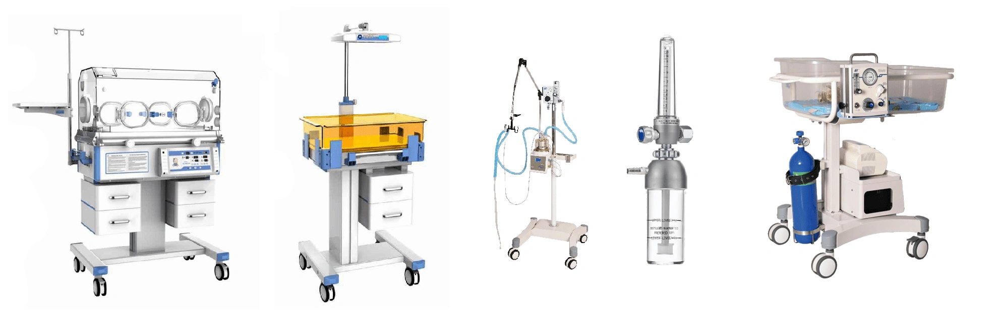
Thermal Regulation Systems: The Shield Against Hypothermia
Premature and low-birth-weight (LBW) infants lack the natural ability to regulate their own body temperature. Without adequate subcutaneous fat layers, they are highly susceptible to cold stress—a dangerous condition that can trigger metabolic complications and organ failure. Consequently, thermal regulation devices serve as the first line of defense in the NICU.
Infant Incubators
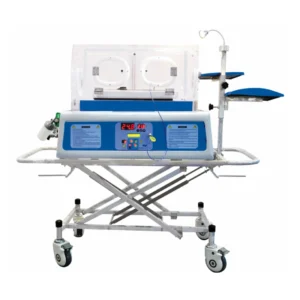
Incubators serve as the primary sanctuary for premature infants. These enclosed, transparent rigid boxes are engineered to maintain a sterile, warm microenvironment with strictly controlled humidity levels. High humidity is critical to prevent trans-epidermal water loss through the infant’s exceptionally thin skin. Furthermore, the incubator’s walls act as a protective barrier against airborne bacterial infections while dampening external hospital noise and harsh light.
Radiant Warmers
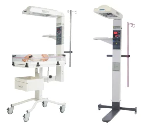
Unlike closed incubators, a radiant warmer is an open-bed system equipped with an overhead infrared heating element. This apparatus is typically utilized in delivery rooms immediately following birth, during emergency resuscitation, or whenever clinicians require rapid, unhindered physical access to the infant for medical procedures. Because the bed remains open, medical staff can move freely without being restricted by canopy walls, while ensuring the baby receives a constant, stabilizing flow of overhead heat.
Servo Heaters
Safety in temperature management is further elevated through the integration of servo-control technology. These smart heating systems connect directly to a sensitive skin temperature probe attached to the infant’s abdomen. The mechanism operates automatically: if the probe detects a drop in the infant’s body temperature, the heating unit increases its output. Conversely, once the optimal body temperature is reached, the heater scales back. This continuous feedback loop eliminates the risk of overheating, which is equally hazardous to a newborn’s safety.
Respiratory Support Devices: Delivering the Breath of Life
The lungs are among the very last organs to mature fully during fetal development. As a result, many neonatal patients suffer from respiratory distress syndrome due to a lack of surfactant—the natural substance that keeps the lung’s air sacs open. To overcome this physiological limitation, the NICU relies on a spectrum of respiratory support machinery, ranging from invasive to non-invasive methods.
Mechanical Ventilators
When a newborn is entirely unable to breathe independently or suffers from severe respiratory failure, a mechanical ventilator becomes the most critical life-sustaining tool. This device operates invasively by delivering warmed, humidified air and oxygen directly into the lungs via an endotracheal tube placed down the infant’s windpipe. Modern neonatal ventilators possess an extraordinary level of precision, capable of delivering minute volumes of air at incredibly gentle pressures to prevent barotrauma to fragile lung tissues.
CPAP (Continuous Positive Airway Pressure) Machines
As a safer, non-invasive alternative for mild to moderate respiratory distress, CPAP machines are frequently the preferred choice. The device delivers a continuous stream of pressurized air and oxygen through small prongs placed inside the infant’s nostrils. This constant positive pressure keeps the alveoli (air sacs) inflated even when the infant exhales, preventing lung collapse and significantly reducing the energy the baby needs to expend for subsequent breaths.
High-Flow Nasal Cannula (HFNC) & Bedside Suction Units
HFNC technology is utilized to deliver heated and humidified oxygen-air blends at higher flow rates than standard cannulas, offering greater comfort for infants transitioning away from heavy mechanical ventilation toward independent breathing. This setup is always paired with a bedside suction unit. Suction devices are vital for clearing mucus, amniotic fluid, or secretions that obstruct the infant’s airway, ensuring a clear path for oxygen without delay.
Monitoring and Therapeutics: Continuous Vigilance and Precise Interventions
The clinical status of a neonatal patient can fluctuate drastically within seconds. Therefore, highly sensitive monitoring systems must be paired with precise therapeutic devices to ensure timely and accurate medical responses.
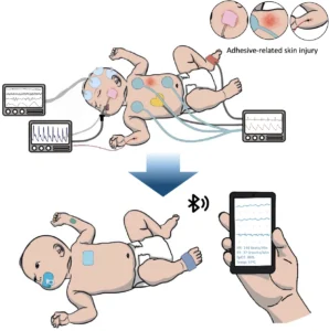
Patient Monitors
Every infant in the NICU is connected to an array of non-invasive sensors linked to a central patient monitor. The display provides real-time tracking of vital signs, including heart rate, respiratory rate, blood pressure, and blood oxygen saturation () via pulse oximetry probes. These systems feature highly sensitive, customized alarm thresholds that instantly alert nursing staff to any sudden drop or spike in vital parameters, enabling immediate clinical intervention.
Phototherapy Units (“Bili Lights”)
Neonatal jaundice, caused by hyperbilirubinemia, is a common condition resulting from an immature, underfunctioning liver. Phototherapy units emit a targeted spectrum of blue light over the infant’s exposed skin. This light breaks down excess bilirubin into a water-soluble form that can be easily excreted through urine and stool. Throughout this therapy, the infant’s eyes and genitals are shielded with specialized protective pads to prevent tissue damage from prolonged light exposure.
Infusion & Syringe Pumps
Premature infants require intravenous fluids, total parenteral nutrition (TPN), and critical medications in exceptionally microscopic, precise doses. Conventional intravenous lines cannot be trusted due to the catastrophic risk of fluid overload. Infusion and syringe pumps solve this challenge by allowing clinical staff to program digital delivery rates down to fractions of a milliliter per hour, ensuring absolute dosing accuracy.
Resuscitation and Emergency Gear: Rapid Action in Critical Moments
In addition to continuous maintenance equipment, the NICU must always be stocked with immediate-use emergency gear to manage sudden apnea or acute cardiac events.
-
Laryngoscopes and Endotracheal (ET) Tubes: A laryngoscope provides the clinician with direct visualization of the vocal cords, allowing an ET tube to be positioned precisely within the airway during emergency intubation.
-
Self-Inflating Resuscitation Bags: Commonly known as bag-valve-mask units, these manual tools are hand-operated by medical staff to deliver positive pressure breaths during acute resuscitation phases before a baby can be transitioned onto a mechanical ventilator.
Synchronized Technology for a New Generation
The presence of these diverse medical devices underscores that neonatal care is a sophisticated combination of clinical expertise and technological excellence. Without the seamless synchronization of thermal, respiratory, and monitoring devices, the survival rate of critically ill newborns would drop drastically.
For modern hospitals and healthcare providers, investing in the routine maintenance, precise calibration, and technological upgrading of NICU equipment remains absolute. Ensuring that every incubator, ventilator, and syringe pump operates with total accuracy is more than just maintaining hospital compliance—it is a direct commitment to giving the most fragile new lives their very best chance to thrive.
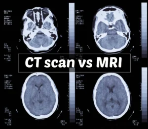
CT Scan vs MRI: Which One Should Hospitals Prioritize?
CT Scan vs MRI: Which One Should Hospitals Prioritize? | In hospital operations, the radiology department serves as the core of diagnostics, determining the accuracy of subsequent medical interventions. Among the advanced devices available, Computed Tomography (CT) Scans and Magnetic Resonance Imaging (MRI) are the two primary instruments clinicians rely on most. While both are designed to non-invasively produce cross-sectional images of the human body, their working principles, technological components, and clinical applications are completely different.
For hospital management, specialists, and biomedical technicians, a deep understanding of this comparative analysis goes beyond basic functionality. It impacts Emergency Room (ER) workflow efficiency, operational cost optimization, and, most importantly, patient safety and diagnostic precision.
Fundamental Tech Principles: Digital X-Rays vs. Magnetic Fields
To understand why these two devices serve different purposes, we must analyze the hardware technology embedded within them.
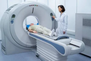
Mechanism of a CT Scan
A CT scan is essentially an advanced evolution of conventional X-ray technology. This device utilizes a motorized rotating X-ray tube inside the gantry (the machine’s tunnel) to encircle the patient’s body. As the tube rotates, detectors capture the X-ray beams that penetrate the body tissues from various angles. This raw data is then processed by a computer using advanced mathematical reconstruction algorithms to produce highly detailed two-dimensional or three-dimensional images of dense anatomical structures.
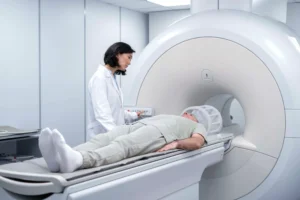
Mechanism of an MRI
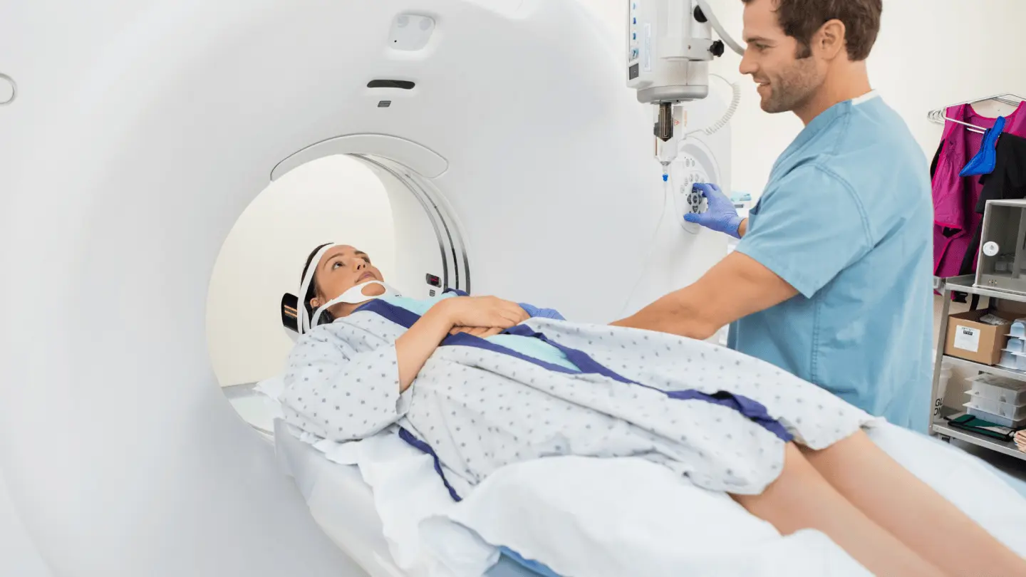
In stark contrast to its counterpart, an MRI does not utilize ionizing radiation. Instead, the machine relies on a high-strength superconducting magnetic field—typically measured in Tesla, such as 1.5T or 3.0T—along with radiofrequency waves.
When a patient enters the MRI bore, the hydrogen atoms within the water molecules of the patient’s body align with the magnetic field. When radiofrequency waves are emitted, this atomic alignment is temporarily disrupted. Once the radio waves are turned off, the atoms return to their original positions while emitting a return radio signal captured by specialized coils. This signal is what the computer converts into images with exceptionally high soft-tissue contrast.
Clinical Emergencies: Why CT Scans Lead the Way in the ER
Time is the most valuable element in the emergency room. When a patient presents with severe trauma or a sudden loss of consciousness, every wasted second can lead to fatal outcomes. In these critical scenarios, CT scan technology demonstrates its absolute dominance.
1. Rapid Evaluation of Trauma and Internal Bleeding
The primary advantage of a CT scan is its imaging speed. The scanning process for the entire body or a specific organ takes only seconds to a few minutes. This characteristic makes it the perfect first-line screening tool to detect internal organ injuries, vascular tears, or internal bleeding from accidents.
2. Acute Stroke Diagnosis
In stroke management, physicians must instantly determine whether symptoms are caused by a blood vessel blockage (ischemic stroke) or a ruptured blood vessel (hemorrhagic stroke). A head CT scan can display blood patches from hemorrhages quickly and clearly, allowing medical teams to make immediate decisions regarding thrombolytic therapy or emergency neurosurgery.
3. Mapping Complex Bone Fractures
While bones can be viewed with standard X-rays, intricate bone structures—such as those in the face, pelvis, or joints experiencing crushed fractures—require the three-dimensional detail provided by a CT scan. The device can visualize the smallest fissures and bone fragment displacements precisely to guide orthopedic surgeons.
4. Chest and Abdomen Examination
CT scans are highly effective for evaluating pathologies within the thoracic and abdominal cavities. Acute disorders such as pulmonary embolism (arterial blockage in the lungs), gastric perforation, ruptured appendix, and bowel obstructions can be identified with high accuracy using iodine-based contrast dyes injected into the bloodstream.
Unmatched Soft Tissue Detail: When Do Clinicians Choose MRI?
While CT scans excel in speed and dense structure visualization, MRI remains the gold standard when physicians require super-detailed imaging of soft, fluid-rich, and hidden components of the body shielded by bone.
1. Diagnosing Complex Neurological Conditions
The brain and spinal cord are highly intricate structures protected by the thick cranium and spinal column. MRI possesses the superior ability to penetrate this bony protection without distortion, sharply distinguishing between healthy and abnormal tissue. Degenerative diseases like multiple sclerosis, micro-sized brain tumors, ultra-early-stage strokes, and spinal cord nerve disorders can only be mapped in detail through MRI scanning.
2. Joint, Muscle, and Ligament Injuries
For athletes or patients suffering from severe sports injuries, an MRI is an indispensable diagnostic tool. The device can reveal ligament tears (such as an ACL tear in the knee), cartilage damage (meniscus), tendon tears, and muscle contusions that would not show up clearly on a standard CT scan or X-ray.
3. Detailed Organ and Pelvic Imaging
MRI provides exceptional visual contrast for evaluating specific internal organs, such as detecting focal lesions in the liver, evaluating cardiac structures, and mapping reproductive organs deep within the pelvic cavity. This significantly assists oncologists in determining accurate cancer staging before surgical interventions.
Technical Considerations and Operational Limitations
Choosing the right device also involves analyzing physical machine limitations, patient comorbidities, and radiation safety aspects.
Risk Factors and Limitations of CT Scans
-
Radiation Exposure: CT scans use ionizing radiation. Although modern medical technology has minimized doses to safe levels, this examination is generally avoided for pregnant patients unless in life-threatening situations. Repeated use on children must also be carefully weighed.
-
Contrast Dye Sensitivity: To enhance vascular structures, CT scans often require an injection of iodine-based contrast media. This agent can trigger allergic reactions in some patients and poses a risk of side effects for individuals with compromised kidney function.
Risk Factors and Limitations of MRIs
-
Duration and Patient Cooperation: An MRI scan takes a significant amount of time, ranging from 20 to 90 minutes depending on the complexity of the targeted area. During this window, patients must remain completely still inside a narrow tunnel. This environment frequently triggers intense anxiety in patients suffering from claustrophobia. Additionally, the loud tapping noises generated by the magnetic coils during operation require patients to wear hearing protection.
-
Metal Hazards and Implantable Devices: Because it uses a powerful magnetic field, the MRI room must remain free of ferromagnetic materials. External metallic objects can be pulled toward the machine at high speeds, posing a severe hazard. For patients with internal medical implants—such as older pacemakers, brain aneurysm clips, or cochlear implants—conventional MRI scans can be highly dangerous, as the magnetic field can displace the implant or disrupt electronic circuitry.
Clinical Decision-Making Guide for Hospitals
To streamline the patient triaging process in radiology departments, the choice of equipment can be summarized based on the following clinical parameters:
-
Choose a CT Scan if: The patient’s condition is an emergency (trauma, accident, suspected acute stroke), the target of the examination is bone structure or the lungs, the patient cannot remain still for long periods (e.g., pediatric or agitated patients), and time efficiency is the top priority.
-
Choose an MRI if: The case is non-emergent but requires specific diagnostics in soft tissues, the examination involves the central nervous system (brain and spine), early tumor detection is needed, chronic ligament injuries are being evaluated, and when the patient must avoid radiation exposure entirely.
By placing patients in the correct imaging modality from the start, hospitals not only save patients from unnecessary diagnostic expenses but also maximize medical equipment utilization according to its best capacity—ensuring excellent, safe, and premium healthcare delivery.
What Is IoMT Security? Protecting Smart Medical Devices
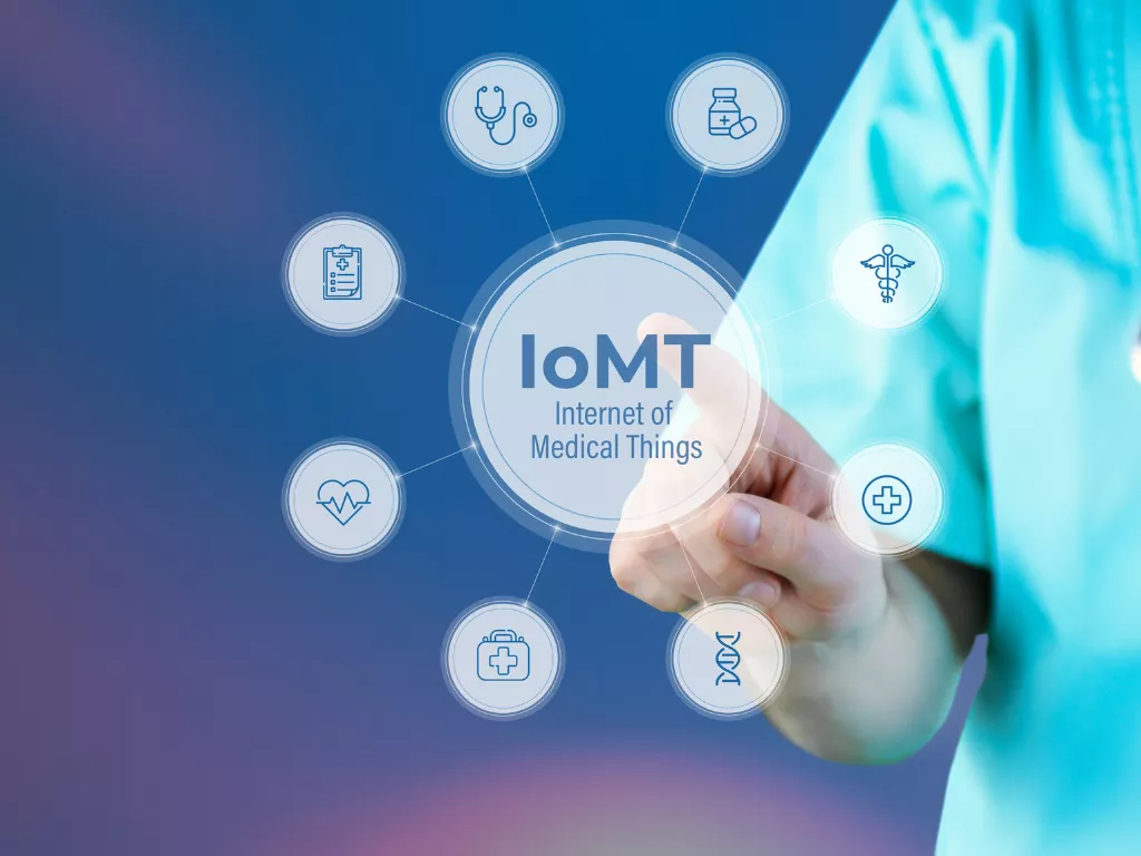
What Is IoMT Security? Protecting Smart Medical Devices | The integration of digital technology into the healthcare sector is advancing at a rapid pace. One of the most significant leaps currently taking place is the adoption of the Internet of Medical Things (IoMT)—a dedicated network connecting various medical devices directly to clinical ecosystems. From implantable pacemakers and smart infusion pumps to advanced hospital imaging scanners, these devices now exchange critical data in real time.
While this interconnectivity offers unprecedented efficiency and diagnostic accuracy, it introduces a critical challenge: ensuring robust security. This is where IoMT security practices become definitive, serving to protect both patient safety and the operational integrity of medical institutions.
Understanding IoMT and Its Scope
In simple terms, the IoMT is an ecosystem of smart medical devices capable of collecting, transmitting, and processing health data over internet networks or local hospital servers. The presence of this technology bridges the gap between clinicians and patients while automating electronic health records (EHR) documentation.
Generally, IoMT devices fall into a few primary categories:
-
Wearable Devices: Portable heart rate monitors or continuous glucose sensors used by patients in their daily lives.
-
Implantable Devices: Medical hardware embedded inside the body, such as modern pacemakers that remotely transmit performance reports to cardiologists.
-
Smart Hospital Facilities: Large-scale equipment including MRI machines, CT scanners, and interconnected smart beds linked to a central clinical dashboard.
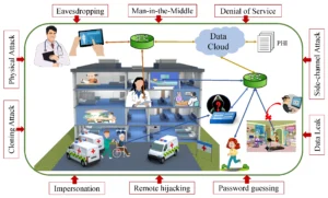
Why Protecting IoMT Systems is a Top Priority
Securing an IoMT network requires a fundamentally different approach than protecting standard IT systems like corporate laptops or smartphones. Securing a medical device directly translates to safeguarding human life. If an IoMT network is breached, the resulting disruption can cause severe chaos within clinical workflows.
There are three primary reasons why protecting IoMT networks is urgent:
1. Patient Safety and Data Protection
Protected Health Information (PHI) is a highly valuable commodity and a frequent target for cybercriminals. While data breaches heavily compromise patient privacy, the unauthorized manipulation of device functionality poses a far greater danger. If a hacker alters the dosage on an infusion pump or silences a cardiac monitor alarm, the patient’s physical safety is immediately jeopardized.
2. The Vulnerability of Legacy Devices
Hospitals frequently rely on high-cost medical equipment purchased years ago. These legacy devices were engineered purely for clinical performance, not to withstand modern cyber attacks. Consequently, many of these devices run on unpatchable firmware and lack the processing memory required to support advanced encryption protocols.
3. New Gaps Introduced by Artificial Intelligence (AI)
Integrating AI to process IoMT diagnostic data yields remarkable efficiency, but it also creates new attack vectors. Sophisticated cyber threats can manipulate data within machine learning models to deceive early-detection systems. Because these subtle attacks bypass conventional IT defense methods, they can quietly distort diagnostic outcomes and mislead physicians.
Strategic Steps to Fortify IoMT Security
To manage a complex, multi-vendor device ecosystem, hospital administrations and biomedical technicians must implement a layered security approach.
-
Network Segmentation: A crucial first step involves isolating the hospital’s public Wi-Fi network (used by visitors) from the internal network dedicated strictly to IoMT hardware. This prevents attackers from accessing medical devices via public entry points.
-
Automated Traffic Monitoring: Utilizing artificial intelligence to continuously monitor data traffic. If an anomaly is detected—such as a pacemaker attempting to transmit data to an unfamiliar external server—the system can automatically block the connection.
-
Clinical Staff Education: Cybersecurity is not solely the responsibility of the IT department. Nurses, doctors, and clinical staff must be trained to recognize operational anomalies in the devices they use daily.
Securing the IoMT is not about restricting innovation. Rather, it is about building a resilient foundation so that future medical technologies can be adopted safely, ultimately elevating the standard of patient care.