
MRI vs CT Scan: Differences, Uses, and Costs Explained
MRI vs CT Scan: Differences, Uses, and Costs Explained | Medical imaging technology has transformed the way doctors diagnose and treat diseases. Two of the most commonly used imaging tests are MRI and CT scans. Both provide detailed images of the inside of the body, but they work differently and are used for different medical situations.
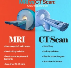
Understanding MRI vs CT Scan: Differences, Uses, and Costs can help patients feel more prepared before undergoing a diagnostic test. While an MRI uses powerful magnets and radio waves to create detailed images, a CT scan uses X-rays combined with computer technology to produce cross-sectional images of the body.
The choice between an MRI and CT scan depends on several factors, including the body area being examined, the medical condition being investigated, urgency, cost, and patient safety considerations.
In this guide, we will explain the key differences between MRI and CT scans, their common uses, advantages, disadvantages, and average costs.
What Is an MRI Scan?
MRI stands for Magnetic Resonance Imaging. It is an advanced medical imaging procedure that uses strong magnetic fields and radio waves to create highly detailed images of organs, tissues, muscles, and other structures inside the body.
Unlike CT scans, MRI does not use ionizing radiation. This makes MRI a preferred option for certain patients who need repeated imaging examinations.
During an MRI scan, the patient lies inside a large tube-shaped machine. The magnetic field temporarily changes the alignment of hydrogen atoms in the body. Radio waves are then used to create detailed images based on how these atoms respond.
MRI technology is especially effective for examining soft tissues that may not appear clearly on other imaging tests.
What Is a CT Scan?
A CT scan, also called a Computed Tomography scan, is an imaging test that combines multiple X-ray images to create detailed cross-sectional views of the body.
Unlike traditional X-rays, which provide a flat image, CT scans create layered images that allow doctors to see structures from different angles.
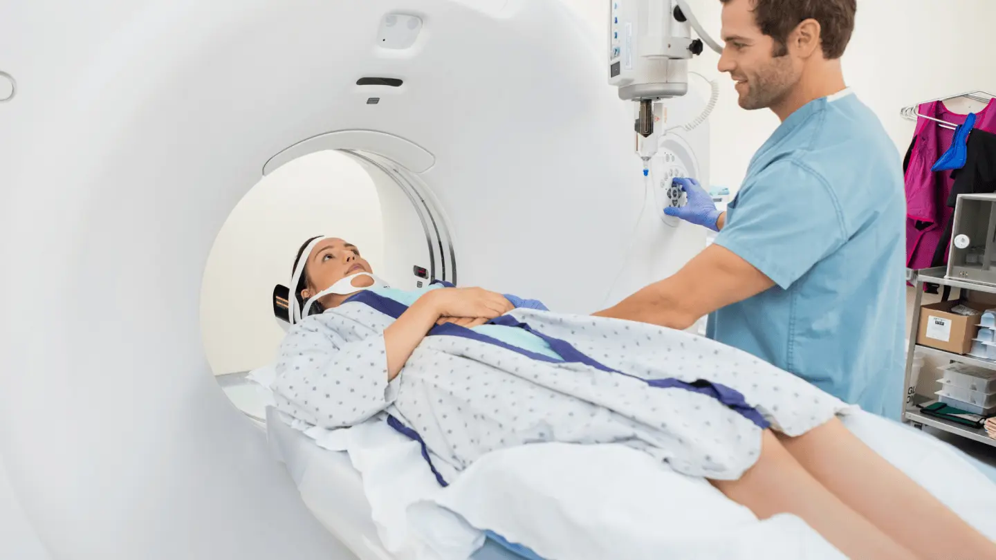
A CT scanner looks like a large circular machine. During the procedure, the patient lies on a table that moves through the scanner while X-ray detectors capture images.
CT scans are widely used because they are fast, reliable, and effective for diagnosing many emergency conditions.
MRI vs CT Scan: Key Differences
Understanding the differences between MRI and CT scans can help explain why doctors choose one test over another.
| Feature | MRI Scan | CT Scan |
|---|---|---|
| Technology | Magnetic fields and radio waves | X-rays and computer processing |
| Radiation Exposure | No ionizing radiation | Uses radiation |
| Scan Time | Usually longer | Usually faster |
| Best For | Soft tissues, brain, nerves, joints | Bones, injuries, emergencies |
| Image Detail | Excellent soft tissue detail | Excellent overall body imaging |
| Cost | Usually more expensive | Usually less expensive |
| Noise Level | Loud | Quieter |
| Emergency Use | Less common | Frequently used |
Both imaging methods are valuable, but they serve different purposes.
MRI vs CT Scan: Which One Provides Better Images?
There is no single answer to which imaging test is better because each technology has different strengths.
MRI Provides Better Soft Tissue Images
MRI is usually preferred for examining:
- Brain structures
- Spinal cord
- Ligaments
- Tendons
- Muscles
- Cartilage
- Internal organs
For example, doctors often use MRI scans to evaluate:
- Brain tumors
- Multiple sclerosis
- Spinal injuries
- Joint problems
- Soft tissue abnormalities
MRI creates extremely detailed images of soft tissues, making it one of the best tools for neurological and musculoskeletal evaluations.
CT Provides Faster Overall Imaging
CT scans are commonly preferred when doctors need quick answers, especially during emergencies.
CT scans are often used for:
- Bone fractures
- Internal bleeding
- Traumatic injuries
- Lung problems
- Abdominal conditions
- Stroke evaluation
Because CT scans can usually be completed within minutes, they are frequently used in emergency rooms.
MRI Uses: When Doctors Recommend an MRI Scan
MRI scans are commonly used for diagnosing conditions involving soft tissues and complex body structures.
Brain and Nervous System Imaging
Doctors may recommend an MRI to examine:
- Brain tumors
- Stroke damage
- Multiple sclerosis
- Brain infections
- Nerve disorders
MRI provides detailed images of brain tissue that help doctors identify abnormalities.
Spine and Back Problems
MRI is often used to diagnose:
- Herniated discs
- Spinal cord injuries
- Nerve compression
- Degenerative spine conditions
Because MRI provides excellent images of discs and nerves, it is often considered the preferred imaging method for many spinal conditions.
Joint and Sports Injuries
Athletes frequently undergo MRI scans to evaluate:
- Torn ligaments
- Muscle injuries
- Cartilage damage
- Tendon problems
MRI can identify injuries that may not appear on standard X-rays.
CT Scan Uses: When Doctors Recommend a CT Scan
CT scans are commonly selected when speed and detailed structural information are important.
Emergency Injuries
CT scans are frequently used after accidents or serious injuries because they quickly detect:
- Internal bleeding
- Bone fractures
- Organ damage
- Head trauma
Fast results can help doctors make immediate treatment decisions.
Lung and Chest Imaging
CT scans can provide detailed images of the lungs and chest area.
Doctors may use CT scans to evaluate:
- Lung nodules
- Pulmonary conditions
- Chest injuries
- Blood vessel problems
Abdominal and Pelvic Conditions
CT scans are commonly used to investigate:
- Kidney stones
- Appendicitis
- Abdominal injuries
- Tumors
- Infections
MRI vs CT Scan Costs
The cost of MRI and CT scans varies depending on several factors, including:
- Location
- Healthcare provider
- Insurance coverage
- Body area being examined
- Use of contrast dye
- Type of imaging equipment
Average MRI Scan Cost
MRI scans are generally more expensive because the equipment is more complex and the procedure often takes longer.
Typical MRI costs may range from:
- $400 to $3,500 or more without insurance
Specialized MRI exams may cost more depending on complexity.
Average CT Scan Cost
CT scans are generally less expensive compared with MRI.
Typical CT scan costs may range from:
- $300 to $3,000 without insurance
Emergency CT scans may have additional hospital-related charges.
Patients should always confirm costs with their healthcare provider and insurance company before scheduling an examination.
MRI vs CT Scan: Safety Differences
Both MRI and CT scans are considered safe when performed correctly, but they have different safety considerations.
MRI Safety Considerations
MRI does not use radiation, but the powerful magnetic field means certain patients must take precautions.
Patients should inform medical staff if they have:
- Metal implants
- Pacemakers
- Certain medical devices
- Metal fragments in the body
MRI machines can also be uncomfortable for people who experience claustrophobia.
CT Scan Safety Considerations
CT scans use X-rays, which involve radiation exposure.
Doctors consider the benefits and risks before recommending CT imaging, especially for:
- Children
- Pregnant patients
- People requiring frequent scans
In many situations, the diagnostic benefits of CT scanning outweigh the potential risks.
MRI vs CT Scan With Contrast Dye
Some MRI and CT scans use contrast agents to improve image clarity.
MRI Contrast
MRI contrast usually contains gadolinium-based substances.
It helps highlight:
- Tumors
- Blood vessels
- Inflammation
- Abnormal tissue
CT Contrast
CT contrast commonly uses iodine-based substances.
It helps doctors examine:
- Blood vessels
- Organs
- Infections
- Tumors
Patients should inform their doctor about allergies or kidney problems before receiving contrast.
MRI or CT Scan: Which One Is Better?
The answer depends on the medical situation.
MRI may be better when doctors need:
- Detailed soft tissue images
- Brain evaluation
- Spine examination
- Joint assessment
CT may be better when doctors need:
- Fast results
- Emergency diagnosis
- Bone imaging
- Trauma evaluation
Doctors choose the imaging method based on accuracy, safety, urgency, and the condition being investigated.
Frequently Asked Questions About MRI vs CT Scan
Is MRI safer than CT scan?
MRI does not use radiation, while CT scans use X-rays. However, both tests are considered safe when medically necessary.
Is MRI more expensive than CT scan?
Yes. MRI scans are usually more expensive because they require advanced equipment and longer scanning times.
How long does an MRI scan take?
Most MRI scans take between 20 and 60 minutes depending on the body area being examined.
How long does a CT scan take?
Many CT scans can be completed within a few minutes.
Can MRI detect cancer?
MRI can help identify tumors and abnormal tissues, but additional tests may be needed to confirm a cancer diagnosis.
Can CT scans detect tumors?
Yes, CT scans can detect many tumors and help doctors evaluate their size and location.
When comparing MRI vs CT Scan: Differences, Uses, and Costs, both imaging technologies provide valuable information for diagnosing medical conditions.
MRI is often the preferred choice for detailed soft tissue imaging, brain examinations, spine problems, and joint injuries. CT scans are commonly chosen for emergencies, trauma, bone injuries, and situations requiring fast results.
The best imaging test depends on your specific medical needs. Your doctor will consider your symptoms, medical history, safety factors, and diagnostic goals before recommending an MRI or CT scan.
Understanding the differences between these two technologies can help you make informed decisions and feel more confident about your healthcare journey.
CT Scan vs MRI: Which One Should Hospitals Prioritize?
CT Scan vs MRI: Which One Should Hospitals Prioritize? | In hospital operations, the radiology department serves as the core of diagnostics, determining the accuracy of subsequent medical interventions. Among the advanced devices available, Computed Tomography (CT) Scans and Magnetic Resonance Imaging (MRI) are the two primary instruments clinicians rely on most. While both are designed to non-invasively produce cross-sectional images of the human body, their working principles, technological components, and clinical applications are completely different.
For hospital management, specialists, and biomedical technicians, a deep understanding of this comparative analysis goes beyond basic functionality. It impacts Emergency Room (ER) workflow efficiency, operational cost optimization, and, most importantly, patient safety and diagnostic precision.
Fundamental Tech Principles: Digital X-Rays vs. Magnetic Fields
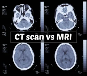
To understand why these two devices serve different purposes, we must analyze the hardware technology embedded within them.
Mechanism of a CT Scan
A CT scan is essentially an advanced evolution of conventional X-ray technology. This device utilizes a motorized rotating X-ray tube inside the gantry (the machine’s tunnel) to encircle the patient’s body. As the tube rotates, detectors capture the X-ray beams that penetrate the body tissues from various angles. This raw data is then processed by a computer using advanced mathematical reconstruction algorithms to produce highly detailed two-dimensional or three-dimensional images of dense anatomical structures.
Mechanism of an MRI
In stark contrast to its counterpart, an MRI does not utilize ionizing radiation. Instead, the machine relies on a high-strength superconducting magnetic field—typically measured in Tesla, such as 1.5T or 3.0T—along with radiofrequency waves.
When a patient enters the MRI bore, the hydrogen atoms within the water molecules of the patient’s body align with the magnetic field. When radiofrequency waves are emitted, this atomic alignment is temporarily disrupted. Once the radio waves are turned off, the atoms return to their original positions while emitting a return radio signal captured by specialized coils. This signal is what the computer converts into images with exceptionally high soft-tissue contrast.
Clinical Emergencies: Why CT Scans Lead the Way in the ER
Time is the most valuable element in the emergency room. When a patient presents with severe trauma or a sudden loss of consciousness, every wasted second can lead to fatal outcomes. In these critical scenarios, CT scan technology demonstrates its absolute dominance.
1. Rapid Evaluation of Trauma and Internal Bleeding
The primary advantage of a CT scan is its imaging speed. The scanning process for the entire body or a specific organ takes only seconds to a few minutes. This characteristic makes it the perfect first-line screening tool to detect internal organ injuries, vascular tears, or internal bleeding from accidents.
2. Acute Stroke Diagnosis
In stroke management, physicians must instantly determine whether symptoms are caused by a blood vessel blockage (ischemic stroke) or a ruptured blood vessel (hemorrhagic stroke). A head CT scan can display blood patches from hemorrhages quickly and clearly, allowing medical teams to make immediate decisions regarding thrombolytic therapy or emergency neurosurgery.
3. Mapping Complex Bone Fractures
While bones can be viewed with standard X-rays, intricate bone structures—such as those in the face, pelvis, or joints experiencing crushed fractures—require the three-dimensional detail provided by a CT scan. The device can visualize the smallest fissures and bone fragment displacements precisely to guide orthopedic surgeons.
4. Chest and Abdomen Examination
CT scans are highly effective for evaluating pathologies within the thoracic and abdominal cavities. Acute disorders such as pulmonary embolism (arterial blockage in the lungs), gastric perforation, ruptured appendix, and bowel obstructions can be identified with high accuracy using iodine-based contrast dyes injected into the bloodstream.
Unmatched Soft Tissue Detail: When Do Clinicians Choose MRI?
While CT scans excel in speed and dense structure visualization, MRI remains the gold standard when physicians require super-detailed imaging of soft, fluid-rich, and hidden components of the body shielded by bone.
1. Diagnosing Complex Neurological Conditions
The brain and spinal cord are highly intricate structures protected by the thick cranium and spinal column. MRI possesses the superior ability to penetrate this bony protection without distortion, sharply distinguishing between healthy and abnormal tissue. Degenerative diseases like multiple sclerosis, micro-sized brain tumors, ultra-early-stage strokes, and spinal cord nerve disorders can only be mapped in detail through MRI scanning.
2. Joint, Muscle, and Ligament Injuries
For athletes or patients suffering from severe sports injuries, an MRI is an indispensable diagnostic tool. The device can reveal ligament tears (such as an ACL tear in the knee), cartilage damage (meniscus), tendon tears, and muscle contusions that would not show up clearly on a standard CT scan or X-ray.
3. Detailed Organ and Pelvic Imaging
MRI provides exceptional visual contrast for evaluating specific internal organs, such as detecting focal lesions in the liver, evaluating cardiac structures, and mapping reproductive organs deep within the pelvic cavity. This significantly assists oncologists in determining accurate cancer staging before surgical interventions.
Technical Considerations and Operational Limitations
Choosing the right device also involves analyzing physical machine limitations, patient comorbidities, and radiation safety aspects.
Risk Factors and Limitations of CT Scans
-
Radiation Exposure: CT scans use ionizing radiation. Although modern medical technology has minimized doses to safe levels, this examination is generally avoided for pregnant patients unless in life-threatening situations. Repeated use on children must also be carefully weighed.
-
Contrast Dye Sensitivity: To enhance vascular structures, CT scans often require an injection of iodine-based contrast media. This agent can trigger allergic reactions in some patients and poses a risk of side effects for individuals with compromised kidney function.
Risk Factors and Limitations of MRIs
-
Duration and Patient Cooperation: An MRI scan takes a significant amount of time, ranging from 20 to 90 minutes depending on the complexity of the targeted area. During this window, patients must remain completely still inside a narrow tunnel. This environment frequently triggers intense anxiety in patients suffering from claustrophobia. Additionally, the loud tapping noises generated by the magnetic coils during operation require patients to wear hearing protection.
-
Metal Hazards and Implantable Devices: Because it uses a powerful magnetic field, the MRI room must remain free of ferromagnetic materials. External metallic objects can be pulled toward the machine at high speeds, posing a severe hazard. For patients with internal medical implants—such as older pacemakers, brain aneurysm clips, or cochlear implants—conventional MRI scans can be highly dangerous, as the magnetic field can displace the implant or disrupt electronic circuitry.
Clinical Decision-Making Guide for Hospitals
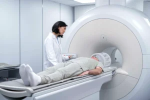
To streamline the patient triaging process in radiology departments, the choice of equipment can be summarized based on the following clinical parameters:
-
Choose a CT Scan if: The patient’s condition is an emergency (trauma, accident, suspected acute stroke), the target of the examination is bone structure or the lungs, the patient cannot remain still for long periods (e.g., pediatric or agitated patients), and time efficiency is the top priority.
-
Choose an MRI if: The case is non-emergent but requires specific diagnostics in soft tissues, the examination involves the central nervous system (brain and spine), early tumor detection is needed, chronic ligament injuries are being evaluated, and when the patient must avoid radiation exposure entirely.
By placing patients in the correct imaging modality from the start, hospitals not only save patients from unnecessary diagnostic expenses but also maximize medical equipment utilization according to its best capacity—ensuring excellent, safe, and premium healthcare delivery.