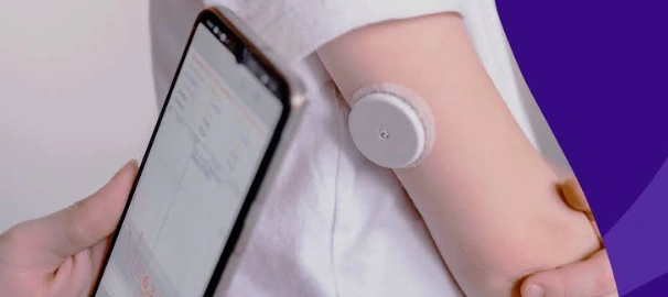
The Future of Glucose Monitoring Dexcom G7 and FreeStyle Libre 3
The Future of Glucose Monitoring Dexcom G7 and FreeStyle Libre 3 | Continuous glucose monitoring (CGM) has changed the way many people track glucose levels. Instead of relying only on occasional finger-stick measurements, CGM systems provide a continuous view of glucose trends throughout the day.

Two of the best-known systems are Dexcom G7 and FreeStyle Libre 3. Both provide real-time glucose information through a wearable sensor, but they differ in areas such as alerts, sensor wear time, data transmission, and device compatibility.
What Is Continuous Glucose Monitoring?
A continuous glucose monitor uses a small wearable sensor to measure glucose levels and provide information through a compatible smartphone, receiver, or other display device.
CGM technology can show not only a current glucose reading but also the direction in which glucose is moving. This can help users identify patterns associated with meals, physical activity, medication, and other daily factors.
However, CGM readings should be interpreted according to the product’s instructions and individual medical needs. If readings do not match symptoms or expectations, users may need to confirm their glucose level with a blood glucose meter before making treatment decisions.
Dexcom G7: Real-Time Monitoring With Advanced Alerts
The Dexcom G7 is designed to provide automatic real-time glucose readings without requiring users to scan the sensor. The system sends glucose information to compatible devices and provides trend information and customizable alerts.
One of its notable features is the Urgent Low Soon alert, which can warn users that glucose is expected to reach a dangerously low level within about 20 minutes. Dexcom also offers customizable high and low alerts and different notification settings.
The standard G7 sensor has a wear time of 10.5 days, including a 12-hour grace period, according to Dexcom’s current product information. It also has a 30-minute warm-up period.
FreeStyle Libre 3: Compact and Frequent Glucose Updates
The FreeStyle Libre 3 is another CGM system designed to provide glucose information continuously through a wearable sensor.
According to a current Dexcom comparison of the systems, FreeStyle Libre 3 sends glucose readings every minute to a compatible display device, while Dexcom G7 sends readings every five minutes. Both systems provide trend information and allow glucose data to be viewed in real time.
The FreeStyle Libre 3 has a listed sensor wear time of 14 days in that comparison. Its warm-up period is listed as 60 minutes.
Dexcom G7 vs FreeStyle Libre 3
Although both systems serve a similar purpose, their features can make them more suitable for different users.
| Feature | Dexcom G7 | FreeStyle Libre 3 |
|---|---|---|
| Glucose monitoring | Real-time | Real-time |
| Automatic readings | Yes | Yes |
| Reading frequency | Every 5 minutes | Every minute |
| Sensor warm-up | 30 minutes | 60 minutes |
| Listed sensor wear | 10.5 days | 14 days |
| Trend information | Yes | Yes |
| High/low alerts | Yes | Yes |
| Predictive low alert | Urgent Low Soon | Availability depends on system/market |
| Data sharing | Yes | Yes |
| Smartphone integration | Compatible devices | Compatible devices |
Features and availability can vary by country, device, age group, and product version, so users should check the manufacturer’s current information before making a decision.
Which System Has Better Alerts?
Alerts are one area where the Dexcom G7 has a particularly strong feature set.
The G7 includes customizable high and low alerts as well as the Urgent Low Soon feature. Users can also customize notification behavior through settings such as quiet modes.
For someone who places a high priority on proactive alerts, these capabilities may make Dexcom G7 attractive.
However, the importance of individual alert features depends on the user’s health needs and treatment plan.
Which One Has a Longer Sensor Life?
Based on the product comparison published by Dexcom, FreeStyle Libre 3 is listed with a 14-day wear period, while the standard Dexcom G7 is listed at 10.5 days including its grace period.
A longer sensor life may be appealing to users who prefer changing their sensor less frequently.
It is also important to distinguish the standard G7 from the newer Dexcom G7 15 Day, which is designed for adults and offers up to 15.5 days of wear including its grace period in the markets where it is available.
Which One Is More Convenient?
Convenience depends on what matters most to the user.
Dexcom G7 offers a short warm-up time and automatic readings without scanning. It can also connect with compatible smartwatches and supports data sharing with caregivers through the Dexcom Follow system.
FreeStyle Libre 3, meanwhile, offers a longer listed sensor life and frequent automatic readings.
For users who want fewer sensor changes, Libre 3 may be appealing. For users who prioritize rapid startup and extensive alert customization, Dexcom G7 may have an advantage.
Accuracy Should Not Be Judged by One Number Alone
Accuracy is naturally an important consideration when comparing CGM systems. However, comparing accuracy requires looking at the specific product version, study population, market, and measurement methodology.
For example, Dexcom’s current provider comparison lists MARD values for Dexcom G7 and FreeStyle Libre 3 Plus, but these figures should not automatically be treated as a direct comparison between the standard G7 and standard Libre 3.
This is an important reminder that product specifications can change as manufacturers introduce new sensor versions.
The Future of Glucose Monitoring
The future of CGM technology appears to be moving toward systems that are smaller, more connected, and more automated.
Dexcom’s newer G7 platform already demonstrates this direction through smartwatch connectivity, remote data sharing, predictive alerts, and integration with other diabetes-management technologies.
At the same time, competition from systems such as FreeStyle Libre continues to encourage improvements in sensor life, data access, connectivity, and ease of use.
The long-term goal is not simply to collect more glucose data. The goal is to make that information easier to understand and more useful for everyday diabetes management.
How to Choose Between Dexcom G7 and FreeStyle Libre 3
There is no single CGM that is automatically best for everyone.
When comparing Dexcom G7 and FreeStyle Libre 3, consider:
- Sensor wear duration
- Warm-up time
- Alert features
- Smartphone compatibility
- Smartwatch connectivity
- Data-sharing options
- Compatibility with insulin-delivery systems
- Availability in your country
- Insurance or out-of-pocket cost
- Recommendations from your healthcare professional
It is also worth checking the exact product version available in your market because features, age indications, compatibility, and availability can differ.
Dexcom G7 vs FreeStyle Libre 3 is not simply a competition between two glucose sensors. Both systems represent the broader evolution of continuous glucose monitoring toward more convenient, connected, and real-time health technology.
Dexcom G7 stands out for its fast warm-up, customizable alerts, predictive low alert, and growing connectivity ecosystem. FreeStyle Libre 3 offers frequent automatic readings and a longer listed sensor wear period.
The right choice ultimately depends on individual needs, medical requirements, device compatibility, availability, and cost. Anyone considering a CGM should discuss the options with a qualified healthcare professional and check the latest product information for their country.

Other Infectious Diseases Monitored by WHO Through Disease Outbreak News and Weekly Epidemiological Reports
Other Infectious Diseases Monitored by WHO Through Disease Outbreak News and Weekly Epidemiological Reports | The spread of infectious diseases remains an important global health
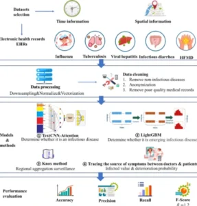 concern. Diseases can cross borders rapidly through travel, migration, trade, environmental changes, and other factors. Because of this, early detection and continuous epidemiological monitoring are essential for protecting communities and reducing the risk of large outbreaks.
The World Health Organization monitors a wide range of infectious diseases through global surveillance systems. Two important sources of epidemiological information are Disease Outbreak News and the Weekly Epidemiological Record.
These publications can highlight newly detected outbreaks, unusual disease events, emerging infections, and developments involving diseases that continue to pose public health risks.
What Is Disease Outbreak News?
Disease Outbreak News is used to communicate important information about significant public health events.
The information may cover an outbreak in a particular country, a cross-border health event, or an emerging infectious disease that requires international attention.
The reports can provide information about:
- The location of an outbreak
- The disease involved
- Reported cases
- Deaths and severe illness
- How the disease is spreading
- Public health measures
- Risk assessments
- Developments in surveillance
The purpose is not to create unnecessary alarm but to help governments, health professionals, and the public understand important developments.
What Is the Weekly Epidemiological Record?
The Weekly Epidemiological Record is a long-running WHO publication dedicated to epidemiological information.
It covers outbreaks and other communicable diseases that have public health importance, including infections that are emerging or returning after periods of lower activity.
Recent weekly reporting has included signals involving diseases such as cholera, mpox, dengue, measles, malaria, yellow fever, Lassa fever, Japanese encephalitis, hantavirus disease, and other infectious threats.
The range of diseases demonstrates that global disease surveillance must cover many different pathogens rather than focusing on only one major outbreak.
Cholera
Cholera remains an important infectious disease monitored internationally.
It is caused by bacteria that can spread through contaminated water or food. Outbreaks are particularly dangerous in areas where access to clean water, sanitation, and healthcare is limited.
Symptoms can include severe watery diarrhea and vomiting. Without rapid treatment, severe dehydration can become life-threatening.
Because cholera can spread quickly within affected communities, early reporting and public health intervention are essential.
Mpox
Mpox continues to receive international surveillance because transmission can occur in different populations and geographic areas.
The disease can cause fever, swollen lymph nodes, fatigue, and characteristic skin lesions. The severity can vary between individuals.
WHO monitoring helps health authorities understand where transmission is occurring and whether changes in epidemiological patterns require additional public health action.
Dengue
Dengue is another major infectious disease monitored globally.
It is primarily transmitted by infected mosquitoes, particularly species of Aedes mosquitoes that are common in tropical and subtropical environments.
Dengue can produce high fever, severe headache, muscle and joint pain, and other symptoms. Some infections can develop into severe dengue, which may require urgent medical treatment.
Urbanization, climate conditions, mosquito populations, and international travel can all influence dengue transmission.
Measles
Measles remains a major concern because it is highly contagious.
The disease can spread rapidly when vaccination coverage falls and susceptible populations increase.
Symptoms commonly include fever, cough, runny nose, and a widespread rash. Complications can be serious, particularly among young children and people with weakened immune systems.
Monitoring measles outbreaks helps public health authorities identify areas where vaccination programs may need to be strengthened.
Malaria
Malaria continues to affect millions of people in tropical and subtropical regions.
It is caused by parasites transmitted through the bites of infected mosquitoes. Although malaria is preventable and treatable, it can become severe and potentially fatal.
WHO surveillance helps track changes in malaria transmission and supports efforts involving prevention, diagnosis, treatment, and vector control.
Yellow Fever
Yellow fever is a mosquito-borne viral disease found in parts of Africa and South America.
Many infections are mild, but severe cases can cause jaundice, bleeding, organ failure, and other life-threatening complications.
Because international travel can move infected people between regions where mosquitoes are present, monitoring yellow fever remains an important part of global health surveillance.
Lassa Fever
Lassa fever is a viral hemorrhagic disease primarily associated with parts of West Africa.
Transmission can occur through exposure to infected rodents or contact with bodily fluids from an infected person.
Many infections may be mild or asymptomatic, but severe cases can produce serious complications.
Regular surveillance is important because early recognition can support infection-control measures and appropriate medical care.
Hantavirus Disease
Hantaviruses are another group of pathogens monitored by health authorities.
Human infection can occur after exposure to environments contaminated with infected rodent urine, droppings, or saliva, depending on the virus involved.
Different hantaviruses can cause different clinical syndromes, including severe respiratory or kidney-related illness.
Because some cases may initially resemble other diseases, epidemiological investigations are important for identifying clusters and potential sources of exposure.
Japanese Encephalitis
Japanese encephalitis is a mosquito-borne viral disease that occurs primarily in parts of Asia and the Western Pacific.
Most infected people do not develop severe symptoms, but a small proportion can develop inflammation of the brain.
Vaccination, mosquito control, and surveillance are important tools for reducing the health impact of the disease.
Zika Virus Disease
Zika virus is mainly transmitted by Aedes mosquitoes.
In many people, infection produces mild symptoms or no symptoms at all. However, Zika is particularly important because infection during pregnancy can cause serious congenital complications.
Monitoring Zika activity is therefore important for both infectious disease control and maternal and child health.
Avian and Animal Influenza Viruses
WHO also monitors influenza viruses that circulate among birds and other animals.
Some animal influenza viruses can occasionally infect humans. When this occurs, health authorities closely examine the circumstances of transmission.
Surveillance is important because influenza viruses can evolve over time. Detecting unusual infections early allows authorities to investigate whether there is evidence of sustained person-to-person transmission.
Viral Hemorrhagic Fevers
Viral hemorrhagic fevers include several diseases caused by different viruses.
Examples include Ebola disease, Marburg virus disease, Lassa fever, and Crimean-Congo hemorrhagic fever.
These diseases vary in their transmission routes and severity, but some can cause serious illness and death.
Rapid detection is particularly important because infection-control measures can help prevent healthcare-associated and community transmission.
Crimean-Congo Hemorrhagic Fever
Crimean-Congo hemorrhagic fever is a tick-borne viral disease that occurs in several regions of the world.
People can become infected through tick bites or contact with infected animal blood and tissues. Human-to-human transmission can also occur through exposure to infected bodily fluids.
Healthcare workers can face particular risks when treating patients without appropriate infection-prevention measures.
Why Weekly Epidemiological Monitoring Matters
Disease surveillance is not simply about counting cases.
Public health experts also examine patterns and changes.
They may ask:
- Is the number of cases increasing?
- Is transmission occurring in a new location?
- Are unusual symptoms being reported?
- Are severe cases becoming more common?
- Is a disease appearing in a population that was previously unaffected?
- Is there evidence of international spread?
- Are existing prevention measures working?
Answers to these questions help authorities decide whether additional investigations or interventions are necessary.
Emerging and Re-Emerging Diseases
An important part of modern disease surveillance is recognizing infections that are emerging or re-emerging.
An emerging disease may involve a pathogen that has newly appeared in humans or is expanding into a new geographic area.
A re-emerging disease may be an infection that was previously under control but begins increasing again.
Changes in climate, environmental conditions, human behavior, urbanization, wildlife interactions, international travel, and healthcare systems can influence these patterns.
The Importance of Early Detection
Early detection can make a major difference during an outbreak.
When health authorities identify an unusual cluster quickly, they may be able to investigate the source, identify contacts, improve infection prevention, provide treatment, and communicate risks before transmission becomes widespread.
This is why global surveillance networks are essential.
A disease event that initially appears to affect only a small community can become an international concern if the pathogen spreads efficiently across borders.
Public Awareness and Responsible Information
Public access to epidemiological information is also important.
However, information about outbreaks should be interpreted carefully.
An infectious disease appearing in a surveillance report does not automatically mean that a global emergency is occurring. Surveillance systems are designed to detect potential threats, including events that may ultimately remain limited.
People should therefore avoid relying on social media rumors or unverified claims when evaluating health risks.
Reliable epidemiological information is most useful when it is presented with appropriate context.
The infectious diseases monitored through WHO surveillance illustrate the complexity of global public health.
Cholera, mpox, dengue, measles, malaria, yellow fever, Lassa fever, hantavirus disease, Japanese encephalitis, Zika virus disease, influenza viruses, and viral hemorrhagic fevers represent only part of the broad range of infections that require continuous observation.
Disease Outbreak News and weekly epidemiological reporting help provide a clearer picture of where unusual health events are occurring and how infectious disease patterns are changing.
The ultimate purpose of this surveillance is early recognition and informed action. By detecting potential threats, assessing their significance, and communicating relevant developments, global health systems can respond more effectively to infectious diseases and reduce their potential impact on communities.

The Medical Tools AI Could Use to Examine Patients
The Medical Tools AI Could Use to Examine Patients | Artificial intelligence is changing the way healthcare professionals collect, analyze, and interpret medical information. As AI becomes more capable, researchers and technology companies are exploring an important possibility: what if AI could do more than analyze data and actually help examine patients?

The answer may involve a combination of AI software, medical sensors, imaging devices, robotic systems, and connected diagnostic tools.
An AI system does not have a physical body by itself. But when connected to the right medical tools, it could potentially gather information about a patient, identify patterns, and support healthcare professionals during clinical examinations.
How Could AI Examine a Patient?
A traditional medical examination involves several steps.
A doctor may ask about symptoms, check vital signs, perform a physical examination, order laboratory tests, review medical history, and analyze imaging results.
AI could potentially assist with many of these steps.
Instead of relying on a single source of information, an AI-powered healthcare system could combine data from multiple medical tools.
For example, it could receive a patient’s heart rate from a wearable sensor, blood oxygen levels from a pulse oximeter, images from an ultrasound device, and laboratory results from a diagnostic system.
The AI could then analyze these different sources together.
1. Digital Stethoscopes
The stethoscope has been one of the most recognizable medical tools for generations.
Digital stethoscopes could give AI systems access to electronically recorded heart and lung sounds.
An AI model could analyze these sounds for patterns that may warrant further investigation.
Potential applications include helping identify abnormal heart sounds or respiratory patterns.
However, an AI-generated interpretation should not automatically be treated as a diagnosis. Medical professionals still need to consider the patient’s symptoms, history, examination, and other test results.
2. Digital Blood Pressure Monitors
Blood pressure is one of the most common measurements taken during a medical examination.
Connected blood pressure monitors could automatically send readings to an AI system.
Instead of looking at a single measurement, AI could potentially analyze trends over time.
For example, repeated readings could help healthcare professionals identify patterns that might otherwise be difficult to recognize from one appointment.
This could be particularly useful for patients who monitor their blood pressure at home.
3. Pulse Oximeters
Pulse oximeters measure blood oxygen saturation and pulse rate.
These small devices are already widely used in healthcare and home monitoring.
When connected to AI, repeated measurements could potentially be analyzed alongside other information such as heart rate, respiratory rate, symptoms, and medical history.
AI could identify changes that deserve attention and potentially alert healthcare professionals.
4. Wearable Health Sensors
Wearable devices could become an important source of continuous health information.
Smartwatches and specialized medical wearables can collect different types of physiological data depending on the device.
Potential measurements can include:
- Heart rate
- Activity levels
- Sleep patterns
- Body temperature
- Heart rhythm information
- Blood oxygen measurements
AI could analyze this information over days or weeks rather than relying only on measurements collected during a short doctor’s appointment.
This could give clinicians a more complete picture of a patient’s health.
5. Ultrasound Devices
Ultrasound is another technology that could become increasingly connected to AI.
Traditional ultrasound examinations require trained professionals to position the probe and interpret the resulting images.
AI could potentially assist by helping identify anatomical structures, improving image acquisition, or highlighting patterns that require further examination.
Portable ultrasound devices could make this technology even more useful in remote or resource-limited environments.
However, ultrasound still requires careful handling and clinical interpretation.
6. X-Ray and Other Medical Imaging Systems
Medical imaging is one of the areas where AI has already demonstrated significant potential.
AI systems can analyze medical images and identify patterns that may be associated with certain diseases or abnormalities.
Tools such as X-ray, CT, MRI, and other imaging systems produce large quantities of information that can be difficult to review manually.
AI could act as an additional layer of analysis.
Instead of replacing a radiologist, the system could highlight areas that deserve closer attention and help prioritize certain cases.
7. Skin Imaging Cameras
AI-powered cameras could potentially help examine visible changes on the skin.
A camera system can capture images of moles, rashes, wounds, or other skin changes.
AI could compare visual characteristics and identify patterns that may warrant professional evaluation.
However, image-based systems can be affected by lighting, skin tone, camera quality, and other factors.
For this reason, AI-based skin analysis should be treated as a support tool rather than a substitute for professional medical evaluation.
8. Digital Otoscopes
An otoscope is used to examine the ear.
Digital otoscopes can capture images or video of the ear canal and eardrum.
AI could potentially analyze these images and help identify patterns associated with common ear conditions.
A connected system could also allow images to be reviewed remotely by healthcare professionals.
This could be particularly useful in areas where access to specialists is limited.
9. Robotic Examination Systems
Robotics could take AI-powered medical examination to another level.
A robotic system equipped with cameras, sensors, and specialized instruments could potentially assist with physical examinations.
In the future, robotic systems might help measure movement, examine specific parts of the body, or perform carefully controlled diagnostic procedures.
However, physical interaction with humans is significantly more complicated than analyzing digital information.
A medical robot would need to operate safely around patients and respond appropriately to unexpected situations.
10. Smart Laboratory Systems
Laboratory testing could also become increasingly integrated with AI.
Blood, urine, and other samples generate large amounts of numerical and biological information.
AI could help identify patterns across multiple laboratory results and compare them with previous measurements.
For example, rather than looking at one laboratory value in isolation, AI could analyze how several values have changed over time.
This could help clinicians identify trends and determine which cases require further investigation.
11. Electronic Health Records
Electronic health records are not physical examination tools, but they could be one of the most important information sources for an AI medical system.
A patient’s record may contain:
- Previous diagnoses
- Medications
- Allergies
- Laboratory results
- Imaging reports
- Clinical notes
- Previous procedures
- Family history
AI could summarize this information and highlight potentially relevant details before a clinical examination.
This could reduce the time clinicians spend searching through records.
12. AI-Powered Cameras and Computer Vision
Computer vision could allow AI systems to analyze visual information about a patient.
Cameras could potentially observe movement, posture, facial expressions, breathing patterns, or other visible characteristics.
In controlled clinical environments, these systems could provide additional information to healthcare professionals.
However, visual information is highly sensitive.
Any system using cameras in healthcare would need strong privacy protections and clear rules about how patient data is collected, stored, and used.
The Power Comes From Combining Tools
The most interesting possibility is not a single AI medical tool.
It is the combination of multiple tools.
Imagine an AI system receiving information from a digital stethoscope, blood pressure monitor, wearable sensor, laboratory system, medical imaging device, and electronic health record.
Individually, each tool provides only part of the picture.
Together, they could create a much richer representation of a patient’s condition.
This is where AI could potentially become especially useful.
AI Still Needs Medical Context
More information does not automatically produce a better diagnosis.
Medical data needs context.
A heart rate of 100 beats per minute may have different meanings depending on whether a person is resting, exercising, anxious, taking medication, or experiencing another medical problem.
An AI system therefore needs to understand not only what the measurement is, but also the circumstances surrounding it.
This is one reason human clinical judgment remains important.
The Importance of Accuracy
Medical AI systems must meet a much higher standard than ordinary consumer software.
A small error in a recommendation can have serious consequences.
For this reason, AI-powered medical tools need appropriate testing, validation, monitoring, and regulatory oversight before being relied upon for clinical decisions.
Healthcare professionals also need to understand the limitations of the technology.
An AI system should not be treated as automatically correct simply because its output looks precise.
Privacy Is Another Major Concern
Medical examination produces highly sensitive information.
Heart data, medical images, laboratory results, genetic information, and clinical records all require strong privacy protections.
As AI systems become more connected, organizations will need to ensure that medical information is collected and processed securely.
Patients should also understand how their information is being used.
Trust will be essential for the adoption of AI-powered healthcare.
Will AI Replace Doctors?
The development of medical examination tools does not necessarily mean doctors will disappear.
In many cases, the more realistic future is AI-assisted medicine.
AI could collect and organize information, identify patterns, monitor patients, and provide decision support.
Doctors could then use that information alongside their own clinical experience and communication skills.
This combination could allow healthcare professionals to spend less time on repetitive data analysis and more time interacting with patients.
The Future of AI-Powered Patient Examination
The future may bring increasingly connected medical environments.
A patient could enter a clinic where sensors automatically collect vital signs. Imaging devices could assist with examination. Laboratory systems could process samples rapidly. AI could combine all of the information and present the clinician with a structured summary.
The doctor would still need to interpret the information and determine the appropriate course of action.
Such a system could make medical care more efficient, but it would also require careful regulation and responsible implementation.
The medical tools AI could use to examine patients range from simple sensors to sophisticated imaging systems and robotic technologies.
Digital stethoscopes, blood pressure monitors, wearable devices, ultrasound machines, medical imaging systems, laboratory equipment, cameras, and electronic health records could all contribute information to an AI-powered healthcare system.
The real potential lies in connecting these tools and allowing AI to analyze information across multiple sources.
But technology should support—not blindly replace—clinical judgment.
The future of healthcare may therefore not be about creating a machine that simply acts like a doctor. It may be about building intelligent medical systems that give doctors better information, help patients receive earlier attention, and make healthcare more efficient while preserving human oversight.

MRI vs CT Scan: Differences, Uses, and Costs Explained
MRI vs CT Scan: Differences, Uses, and Costs Explained | Medical imaging technology has transformed the way doctors diagnose and treat diseases. Two of the most commonly used imaging tests are MRI and CT scans. Both provide detailed images of the inside of the body, but they work differently and are used for different medical situations.
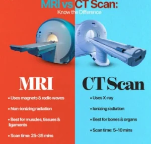
Understanding MRI vs CT Scan: Differences, Uses, and Costs can help patients feel more prepared before undergoing a diagnostic test. While an MRI uses powerful magnets and radio waves to create detailed images, a CT scan uses X-rays combined with computer technology to produce cross-sectional images of the body.
The choice between an MRI and CT scan depends on several factors, including the body area being examined, the medical condition being investigated, urgency, cost, and patient safety considerations.
In this guide, we will explain the key differences between MRI and CT scans, their common uses, advantages, disadvantages, and average costs.
What Is an MRI Scan?
MRI stands for Magnetic Resonance Imaging. It is an advanced medical imaging procedure that uses strong magnetic fields and radio waves to create highly detailed images of organs, tissues, muscles, and other structures inside the body.
Unlike CT scans, MRI does not use ionizing radiation. This makes MRI a preferred option for certain patients who need repeated imaging examinations.
During an MRI scan, the patient lies inside a large tube-shaped machine. The magnetic field temporarily changes the alignment of hydrogen atoms in the body. Radio waves are then used to create detailed images based on how these atoms respond.
MRI technology is especially effective for examining soft tissues that may not appear clearly on other imaging tests.
What Is a CT Scan?
A CT scan, also called a Computed Tomography scan, is an imaging test that combines multiple X-ray images to create detailed cross-sectional views of the body.
Unlike traditional X-rays, which provide a flat image, CT scans create layered images that allow doctors to see structures from different angles.
A CT scanner looks like a large circular machine. During the procedure, the patient lies on a table that moves through the scanner while X-ray detectors capture images.
CT scans are widely used because they are fast, reliable, and effective for diagnosing many emergency conditions.
MRI vs CT Scan: Key Differences
Understanding the differences between MRI and CT scans can help explain why doctors choose one test over another.
| Feature | MRI Scan | CT Scan |
|---|---|---|
| Technology | Magnetic fields and radio waves | X-rays and computer processing |
| Radiation Exposure | No ionizing radiation | Uses radiation |
| Scan Time | Usually longer | Usually faster |
| Best For | Soft tissues, brain, nerves, joints | Bones, injuries, emergencies |
| Image Detail | Excellent soft tissue detail | Excellent overall body imaging |
| Cost | Usually more expensive | Usually less expensive |
| Noise Level | Loud | Quieter |
| Emergency Use | Less common | Frequently used |
Both imaging methods are valuable, but they serve different purposes.
MRI vs CT Scan: Which One Provides Better Images?
There is no single answer to which imaging test is better because each technology has different strengths.
MRI Provides Better Soft Tissue Images
MRI is usually preferred for examining:
- Brain structures
- Spinal cord
- Ligaments
- Tendons
- Muscles
- Cartilage
- Internal organs
For example, doctors often use MRI scans to evaluate:
- Brain tumors
- Multiple sclerosis
- Spinal injuries
- Joint problems
- Soft tissue abnormalities
MRI creates extremely detailed images of soft tissues, making it one of the best tools for neurological and musculoskeletal evaluations.
CT Provides Faster Overall Imaging
CT scans are commonly preferred when doctors need quick answers, especially during emergencies.
CT scans are often used for:
- Bone fractures
- Internal bleeding
- Traumatic injuries
- Lung problems
- Abdominal conditions
- Stroke evaluation
Because CT scans can usually be completed within minutes, they are frequently used in emergency rooms.
MRI Uses: When Doctors Recommend an MRI Scan
MRI scans are commonly used for diagnosing conditions involving soft tissues and complex body structures.
Brain and Nervous System Imaging
Doctors may recommend an MRI to examine:
- Brain tumors
- Stroke damage
- Multiple sclerosis
- Brain infections
- Nerve disorders
MRI provides detailed images of brain tissue that help doctors identify abnormalities.
Spine and Back Problems
MRI is often used to diagnose:
- Herniated discs
- Spinal cord injuries
- Nerve compression
- Degenerative spine conditions
Because MRI provides excellent images of discs and nerves, it is often considered the preferred imaging method for many spinal conditions.
Joint and Sports Injuries
Athletes frequently undergo MRI scans to evaluate:
- Torn ligaments
- Muscle injuries
- Cartilage damage
- Tendon problems
MRI can identify injuries that may not appear on standard X-rays.
CT Scan Uses: When Doctors Recommend a CT Scan
CT scans are commonly selected when speed and detailed structural information are important.
Emergency Injuries
CT scans are frequently used after accidents or serious injuries because they quickly detect:
- Internal bleeding
- Bone fractures
- Organ damage
- Head trauma
Fast results can help doctors make immediate treatment decisions.
Lung and Chest Imaging
CT scans can provide detailed images of the lungs and chest area.
Doctors may use CT scans to evaluate:
- Lung nodules
- Pulmonary conditions
- Chest injuries
- Blood vessel problems
Abdominal and Pelvic Conditions
CT scans are commonly used to investigate:
- Kidney stones
- Appendicitis
- Abdominal injuries
- Tumors
- Infections
MRI vs CT Scan Costs
The cost of MRI and CT scans varies depending on several factors, including:
- Location
- Healthcare provider
- Insurance coverage
- Body area being examined
- Use of contrast dye
- Type of imaging equipment
Average MRI Scan Cost
MRI scans are generally more expensive because the equipment is more complex and the procedure often takes longer.
Typical MRI costs may range from:
- $400 to $3,500 or more without insurance
Specialized MRI exams may cost more depending on complexity.
Average CT Scan Cost
CT scans are generally less expensive compared with MRI.
Typical CT scan costs may range from:
- $300 to $3,000 without insurance
Emergency CT scans may have additional hospital-related charges.
Patients should always confirm costs with their healthcare provider and insurance company before scheduling an examination.
MRI vs CT Scan: Safety Differences
Both MRI and CT scans are considered safe when performed correctly, but they have different safety considerations.
MRI Safety Considerations
MRI does not use radiation, but the powerful magnetic field means certain patients must take precautions.
Patients should inform medical staff if they have:
- Metal implants
- Pacemakers
- Certain medical devices
- Metal fragments in the body
MRI machines can also be uncomfortable for people who experience claustrophobia.
CT Scan Safety Considerations
CT scans use X-rays, which involve radiation exposure.
Doctors consider the benefits and risks before recommending CT imaging, especially for:
- Children
- Pregnant patients
- People requiring frequent scans
In many situations, the diagnostic benefits of CT scanning outweigh the potential risks.
MRI vs CT Scan With Contrast Dye
Some MRI and CT scans use contrast agents to improve image clarity.
MRI Contrast
MRI contrast usually contains gadolinium-based substances.
It helps highlight:
- Tumors
- Blood vessels
- Inflammation
- Abnormal tissue
CT Contrast
CT contrast commonly uses iodine-based substances.
It helps doctors examine:
- Blood vessels
- Organs
- Infections
- Tumors
Patients should inform their doctor about allergies or kidney problems before receiving contrast.
MRI or CT Scan: Which One Is Better?
The answer depends on the medical situation.
MRI may be better when doctors need:
- Detailed soft tissue images
- Brain evaluation
- Spine examination
- Joint assessment
CT may be better when doctors need:
- Fast results
- Emergency diagnosis
- Bone imaging
- Trauma evaluation
Doctors choose the imaging method based on accuracy, safety, urgency, and the condition being investigated.
Frequently Asked Questions About MRI vs CT Scan
Is MRI safer than CT scan?
MRI does not use radiation, while CT scans use X-rays. However, both tests are considered safe when medically necessary.
Is MRI more expensive than CT scan?
Yes. MRI scans are usually more expensive because they require advanced equipment and longer scanning times.
How long does an MRI scan take?
Most MRI scans take between 20 and 60 minutes depending on the body area being examined.
How long does a CT scan take?
Many CT scans can be completed within a few minutes.
Can MRI detect cancer?
MRI can help identify tumors and abnormal tissues, but additional tests may be needed to confirm a cancer diagnosis.
Can CT scans detect tumors?
Yes, CT scans can detect many tumors and help doctors evaluate their size and location.
When comparing MRI vs CT Scan: Differences, Uses, and Costs, both imaging technologies provide valuable information for diagnosing medical conditions.
MRI is often the preferred choice for detailed soft tissue imaging, brain examinations, spine problems, and joint injuries. CT scans are commonly chosen for emergencies, trauma, bone injuries, and situations requiring fast results.
The best imaging test depends on your specific medical needs. Your doctor will consider your symptoms, medical history, safety factors, and diagnostic goals before recommending an MRI or CT scan.
Understanding the differences between these two technologies can help you make informed decisions and feel more confident about your healthcare journey.